Myelin remodeling through experience-dependent oligodendrogenesis in the adult somatosensory cortex.

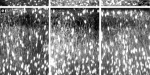
Oligodendrocyte generation in the adult CNS provides a means to adapt the properties of circuits to changes in life experience. However, little is known about the dynamics of oligodendrocytes and the extent of myelin remodeling in the mature brain. Using longitudinal in vivo two-photon imaging of oligodendrocytes and their progenitors in the mouse cerebral cortex, we show that myelination is an inefficient and extended process, with half of the final complement of oligodendrocytes generated after 4 months of age. Oligodendrocytes that successfully integrated formed new sheaths on unmyelinated and sparsely myelinated axons, and they were extremely stable, gradually changing the pattern of myelination. Sensory enrichment robustly increased oligodendrocyte integration, but did not change the length of existing sheaths. This experience-dependent enhancement of myelination in the mature cortex may accelerate information transfer in these circuits and strengthen the ability of axons to sustain activity by providing additional metabolic support.
Oligodendrocytes control potassium accumulation in white matter and seizure susceptibility

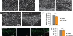
The inwardly rectifying K+ channel Kir4.1 is broadly expressed by CNS glia and deficits in Kir4.1 lead to seizures and myelin vacuolization. However, the role of oligodendrocyte Kir4.1 channels in controlling myelination and K+ clearance in white matter has not been defined. Here, we show that selective deletion of Kir4.1 from oligodendrocyte progenitors (OPCs) or mature oligodendrocytes did not impair their development or disrupt the structure of myelin. However, mice lacking oligodendrocyte Kir4.1 channels exhibited profound functional impairments, including slower clearance of extracellular K+ and delayed recovery of axons from repetitive stimulation in white matter, as well as spontaneous seizures, a lower seizure threshold, and activity-dependent motor deficits. These results indicate that Kir4.1 channels in oligodendrocytes play an important role in extracellular K+ homeostasis in white matter, and that selective loss of this channel from oligodendrocytes is sufficient to impair K+ clearance and promote seizures.

Lineage tracing reveals dynamic changes in oligodendrocyte precursor cells following cuprizone-induced demyelination

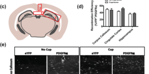
The regeneration of oligodendrocytes is a crucial step in recovery from demyelination, as surviving oligodendrocytes exhibit limited structural plasticity and rarely form additional myelin sheaths. New oligodendrocytes arise through the differentiation of platelet-derived growth factor receptor α (PDGFRα) expressing oligodendrocyte progenitor cells (OPCs) that are widely distributed throughout the CNS. Although there has been detailed investigation of the behavior of these progenitors in white matter, recent studies suggest that disease burden in multiple sclerosis (MS) is more strongly correlated with gray matter atrophy. The timing and efficiency of remyelination in gray matter is distinct from white matter, but the dynamics of OPCs that contribute to these differences have not been defined. Here, we used in vivo genetic fate tracing to determine the behavior of OPCs in gray and white matter regions in response to cuprizone-induced demyelination. Our studies indicate that the temporal dynamics of OPC differentiation varies significantly between white and gray matter. While OPCs rapidly repopulate the corpus callosum and mature into CC1 expressing mature oligodendrocytes, OPC differentiation in the cingulate cortex and hippocampus occurs much more slowly, resulting in a delay in remyelination relative to the corpus callosum. The protracted maturation of OPCs in gray matter may contribute to greater axonal pathology and disease burden in MS.

Changes in the Excitability of Neocortical Neurons in a Mouse Model of Amyotrophic Lateral Sclerosis Are Not Specific to Corticospinal Neurons and Are Modulated by Advancing Disease

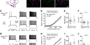
Cell type-specific changes in neuronal excitability have been proposed to contribute to the selective degeneration of corticospinal neurons in amyotrophic lateral sclerosis (ALS) and to neocortical hyperexcitability, a prominent feature of both inherited and sporadic variants of the disease, but the mechanisms underlying selective loss of specific cell types in ALS are not known. We analyzed the physiological properties of distinct classes of cortical neurons in the motor cortex of hSOD1G93A mice of both sexes and found that they all exhibit increases in intrinsic excitability that depend on disease stage. Targeted recordings and in vivo calcium imaging further revealed that neurons adapt their functional properties to normalize cortical excitability as the disease progresses. Although different neuron classes all exhibited increases in intrinsic excitability, transcriptional profiling indicated that the molecular mechanisms underlying these changes are cell type specific. The increases in excitability in both excitatory and inhibitory cortical neurons show that selective dysfunction of neuronal cell types cannot account for the specific vulnerability of corticospinal motor neurons in ALS. Furthermore, the stage-dependent alterations in neuronal function highlight the ability of cortical circuits to adapt as disease progresses. These findings show that both disease stage and cell type must be considered when developing therapeutic strategies for treating ALS.

Myelinogenic Plasticity of Oligodendrocyte Precursor Cells following Spinal Cord Contusion Injury

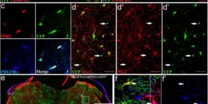
Spontaneous remyelination occurs after spinal cord injury (SCI), but the extent of myelin repair and identity of the cells responsible remain incompletely understood and contentious. We assessed the cellular origin of new myelin by fate mapping platelet-derived growth factor receptor α (PDGFRα), Olig2+, and P0+ cells following contusion SCI in mice. Oligodendrocyte precursor cells (OPCs; PDGFRα+) produced oligodendrocytes responsible for de novo ensheathment of ∼30% of myelinated spinal axons at injury epicenter 3 months after SCI, demonstrating that these resident cells are a major contributor to oligodendrocyte regeneration. OPCs also produced the majority of myelinating Schwann cells in the injured spinal cord; invasion of peripheral myelinating (P0+) Schwann cells made only a limited contribution. These findings reveal that PDGFRα+ cells perform diverse roles in CNS repair, as multipotential progenitors that generate both classes of myelinating cells. This endogenous repair might be exploited as a therapeutic target for CNS trauma and disease.

Cell-type specific differences in promoter activity of the ALS-linked C9orf72 mouse ortholog

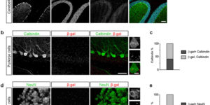
A hexanucleotide repeat expansion in the C9orf72 gene is the most common cause of inherited forms of the neurodegenerative disease amyotrophic lateral sclerosis (ALS). Both loss-of-function and gain-of-function mechanisms have been proposed to underlie this disease, but the pathogenic pathways are not fully understood. To better understand the involvement of different cell types in the pathogenesis of ALS, we systematically analyzed the distribution of promoter activity of the mouse ortholog of C9orf72 in the central nervous system. We demonstrate that C9orf72 promoter activity is widespread in both excitatory and inhibitory neurons as well as in oligodendrocytes and oligodendrocyte precursor cells. In contrast, few microglia and astrocytes exhibit detectable C9orf72 promoter activity. Although at a gross level, the distribution of C9orf72 promoter activity largely follows overall cellular density, we found that it is selectively enriched in subsets of neurons and glial cells that degenerate in ALS. Specifically, we show that C9orf72 promoter activity is enriched in corticospinal and spinal motor neurons as well as in oligodendrocytes in brain regions that are affected in ALS. These results suggest that cell autonomous changes in both neurons and glia may contribute to C9orf72-mediated disease, as has been shown for mutations in superoxide dismutase-1 (SOD1).

Synergistic Signaling by Light and Acetylcholine in Mouse Iris Sphincter Muscle

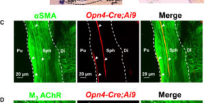
The mammalian pupillary light reflex (PLR) involves a bilateral brain circuit whereby afferent light signals in the optic nerve ultimately drive iris-sphincter-muscle contraction via excitatory cholinergic parasympathetic innervation. Additionally, the PLR in nocturnal and crepuscular sub-primate mammals has a “local” component in the isolated sphincter muscle, as in amphibians, fish, and bird. In mouse, this local PLR requires the pigment melanopsin, originally found in intrinsically photosensitive retinal ganglion cells (ipRGCs). However, melanopsin’s presence and effector pathway locally in the iris remain uncertain. The sphincter muscle itself may express melanopsin, or its cholinergic parasympathetic innervation may be modulated by suggested intraocular axonal collaterals of ipRGCs traveling to the eye’s ciliary body or even to the iris. Here, we show that the muscarinic receptor antagonist, atropine, eliminated the effect of acetylcholine (ACh), but not of light, on isolated mouse sphincter muscle. Conversely, selective genetic deletion of melanopsin in smooth muscle mostly removed the light-induced, but not the ACh-triggered, increase in isolated sphincter muscle’s tension and largely suppressed the local PLR in vivo. Thus, sphincter muscle cells are bona fide, albeit unconventional, photoreceptors. We found melanopsin expression in a small subset of mouse iris sphincter muscle cells, with the light-induced contractile signal apparently spreading through gap junctions into neighboring muscle cells. Light and ACh share a common signaling pathway in sphincter muscle. In summary, our experiments have provided details of a photosignaling process in the eye occurring entirely outside the retina.

Focus scanning with feedback-control for fiber-optic nonlinear endomicroscopy

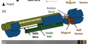
Fiber-optic endomicroscopes open new avenues for the application of non-linear optics to novel in vivo applications. To achieve focus scanning in vivo, shape memory alloy (SMA) wires have been used to move optical elements in miniature endomicroscopes. However, this method has various limitations, making it difficult to achieve accurate and reliable depth scanning. Here we present a feedback-controlled SMA depth scanner. With a Hall effect sensor, contraction of the SMA wire can be tracked in real time, rendering accurate and robust control of motion. The SMA depth scanner can achieve up to 490 µm travel and with open-loop operation, it can move more than 350 µm within one second. With the feedback loop engaged, submicron positioning accuracy was achieved along with superior positioning stability. The high-precision positioning capability of the SMA depth scanner was verified by depth-resolved nonlinear endomicroscopic imaging of mouse brain samples.

Transient Opening of the Mitochondrial Permeability Transition Pore Induces Microdomain Calcium Transients in Astrocyte Processes.

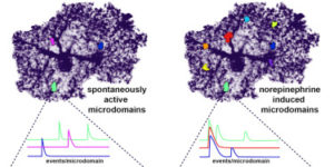
Astrocytes extend highly branched processes that form functionally isolated microdomains, facilitating local homeostasis by redistributing ions, removing neurotransmitters, and releasing factors to influence blood flow and neuronal activity. Microdomains exhibit spontaneous increases in calcium (Ca2+), but the mechanisms and functional significance of this localized signaling are unknown. By developing conditional, membrane-anchored GCaMP3 mice, we found that microdomain activity that occurs in the absence of inositol triphosphate (IP3)-dependent release from endoplasmic reticulum arises through Ca2+ efflux from mitochondria during brief openings of the mitochondrial permeability transition pore. These microdomain Ca2+ transients were facilitated by the production of reactive oxygen species during oxidative phosphorylation and were enhanced by expression of a mutant form of superoxide dismutase 1 (SOD1 G93A) that causes astrocyte dysfunction and neurodegeneration in amyotrophic lateral sclerosis (ALS). By localizing mitochondria to microdomains, astrocytes ensure local metabolic support for energetically demanding processes and enable coupling between metabolic demand and Ca2+ signaling events.

Activity-dependent switch of GABAergic inhibition into glutamatergic excitation in astrocyte-neuron networks.

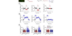
Interneurons are critical for proper neural network function and can activate Ca2+ signaling in astrocytes. However, the impact of the interneuron-astrocyte signaling into neuronal network operation remains unknown. Using the simplest hippocampal Astrocyte-Neuron network, i.e., GABAergic interneuron, pyramidal neuron, single CA3-CA1 glutamatergic synapse, and astrocytes, we found that interneuron-astrocyte signaling dynamically affected excitatory neurotransmission in an activity- and time-dependent manner, and determined the sign (inhibition vs potentiation) of the GABA-mediated effects. While synaptic inhibition was mediated by GABAA receptors, potentiation involved astrocyte GABAB receptors, astrocytic glutamate release, and presynaptic metabotropic glutamate receptors. Using conditional astrocyte-specific GABAB receptor (Gabbr1) knockout mice, we confirmed the glial source of the interneuron-induced potentiation, and demonstrated the involvement of astrocytes in hippocampal theta and gamma oscillations in vivo. Therefore, astrocytes decode interneuron activity and transform inhibitory into excitatory signals, contributing to the emergence of novel network properties resulting from the interneuron-astrocyte interplay.