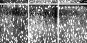
Persistent Cyfip1 Expression Is Required to Maintain the Adult Subventricular Zone Neurogenic Niche

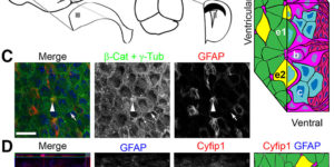
Neural stem cells (NSCs) persist throughout life in the subventricular zone (SVZ) neurogenic niche of the lateral ventricles as Type B1 cells in adult mice. Maintaining this population of NSCs depends on the balance between quiescence and self-renewing or self-depleting cell divisions. Interactions between B1 cells and the surrounding niche are important in regulating this balance, but the mechanisms governing these processes have not been fully elucidated. The cytoplasmic FMRP-interacting protein (Cyfip1) regulates apical-basal polarity in the embryonic brain. Loss of Cyfip1 during embryonic development in mice disrupts the embryonic niche and affects cortical neurogenesis. However, a direct role for Cyfip1 in the regulation of adult NSCs has not been established. Here, we demonstrate that Cyfip1 expression is preferentially localized to B1 cells in the adult mouse SVZ. Loss of Cyfip1 in the embryonic mouse brain results in altered adult SVZ architecture and expansion of the adult B1 cell population at the ventricular surface. Furthermore, acute deletion of Cyfip1 in adult NSCs results in a rapid change in adherens junction proteins as well as increased proliferation and number of B1 cells at the ventricular surface. Together, these data indicate that Cyfip1 plays a critical role in the formation and maintenance of the adult SVZ niche; furthermore, deletion of Cyfip1 unleashes the capacity of adult B1 cells for symmetric renewal to increase the adult NSC pool.
Purinergic Signaling in Cochlear Supporting Cells Reduces Hair Cell Excitability by Increasing the Extracellular Space

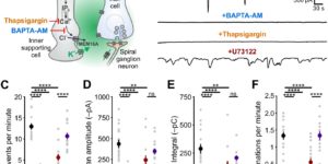
Neurons in developing sensory pathways exhibit spontaneous bursts of electrical activity that are critical for survival, maturation and circuit refinement. In the auditory system, intrinsically generated activity arises within the cochlea, but the molecular mechanisms that initiate this activity remain poorly understood. We show that burst firing of mouse inner hair cells prior to hearing onset requires P2RY1 autoreceptors expressed by inner supporting cells. P2RY1 activation triggers K+ efflux and depolarization of hair cells, as well as osmotic shrinkage of supporting cells that dramatically increased the extracellular space and speed of K+ redistribution. Pharmacological inhibition or genetic disruption of P2RY1 suppressed neuronal burst firing by reducing K+ release, but unexpectedly enhanced their tonic firing, as water resorption by supporting cells reduced the extracellular space, leading to K+ accumulation. These studies indicate that purinergic signaling in supporting cells regulates hair cell excitability by controlling the volume of the extracellular space.

Glutamatergic synaptic input to glioma cells drives brain tumour progression.

A network of communicating tumour cells that is connected by tumour microtubes mediates the progression of incurable gliomas. Moreover, neuronal activity can foster malignant behaviour of glioma cells by non-synaptic paracrine and autocrine mechanisms. Here we report a direct communication channel between neurons and glioma cells in different disease models and human tumours: functional bona fide chemical synapses between presynaptic neurons and postsynaptic glioma cells. These neurogliomal synapses show a typical synaptic ultrastructure, are located on tumour microtubes, and produce postsynaptic currents that are mediated by glutamate receptors of the AMPA subtype. Neuronal activity including epileptic conditions generates synchronised calcium transients in tumour-microtube-connected glioma networks. Glioma-cell-specific genetic perturbation of AMPA receptors reduces calcium-related invasiveness of tumour-microtube-positive tumour cells and glioma growth. Invasion and growth are also reduced by anaesthesia and the AMPA receptor antagonist perampanel, respectively. These findings reveal a biologically relevant direct synaptic communication between neurons and glioma cells with potential clinical implications.

Electrical and synaptic integration of glioma into neural circuits.

High-grade gliomas are lethal brain cancers whose progression is robustly regulated by neuronal activity. Activity-regulated release of growth factors promotes glioma growth, but this alone is insufficient to explain the effect that neuronal activity exerts on glioma progression. Here we show that neuron and glioma interactions include electrochemical communication through bona fide AMPA receptor-dependent neuron-glioma synapses. Neuronal activity also evokes non-synaptic activity-dependent potassium currents that are amplified by gap junction-mediated tumour interconnections, forming an electrically coupled network. Depolarization of glioma membranes assessed by in vivo optogenetics promotes proliferation, whereas pharmacologically or genetically blocking electrochemical signalling inhibits the growth of glioma xenografts and extends mouse survival. Emphasizing the positive feedback mechanisms by which gliomas increase neuronal excitability and thus activity-regulated glioma growth, human intraoperative electrocorticography demonstrates increased cortical excitability in the glioma-infiltrated brain. Together, these findings indicate that synaptic and electrical integration into neural circuits promotes glioma progression.

Oligodendrocyte precursor cells present antigen and are cytotoxic targets in inflammatory demyelination.

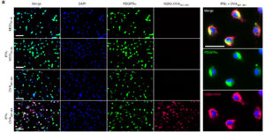
Oligodendrocyte precursor cells (OPCs) are abundant in the adult central nervous system, and have the capacity to regenerate oligodendrocytes and myelin. However, in inflammatory diseases such as multiple sclerosis (MS) remyelination is often incomplete. To investigate how neuroinflammation influences OPCs, we perform in vivo fate-tracing in an inflammatory demyelinating mouse model. Here we report that OPC differentiation is inhibited by both effector T cells and IFNγ overexpression by astrocytes. IFNγ also reduces the absolute number of OPCs and alters remaining OPCs by inducing the immunoproteasome and MHC class I. In vitro, OPCs exposed to IFNγ cross-present antigen to cytotoxic CD8 T cells, resulting in OPC death. In human demyelinated MS brain lesions, but not normal appearing white matter, oligodendroglia exhibit enhanced expression of the immunoproteasome subunit PSMB8. Therefore, OPCs may be co-opted by the immune system in MS to perpetuate the autoimmune response, suggesting that inhibiting immune activation of OPCs may facilitate remyelination.

Molecularly defined cortical astroglia subpopulation modulates neurons via secretion of Norrin.

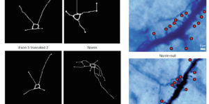
Despite expanding knowledge regarding the role of astroglia in regulating neuronal function, little is known about regional or functional subgroups of brain astroglia and how they may interact with neurons. We use an astroglia-specific promoter fragment in transgenic mice to identify an anatomically defined subset of adult gray matter astroglia. Using transcriptomic and histological analyses, we generate a combinatorial profile for the in vivo identification and characterization of this astroglia subpopulation. These astroglia are enriched in mouse cortical layer V; express distinct molecular markers, including Norrin and leucine-rich repeat-containing G-protein-coupled receptor 6 (LGR6), with corresponding layer-specific neuronal ligands; are found in the human cortex; and modulate neuronal activity. Astrocytic Norrin appears to regulate dendrites and spines; its loss, as occurring in Norrie disease, contributes to cortical dendritic spine loss. These studies provide evidence that human and rodent astroglia subtypes are regionally and functionally distinct, can regulate local neuronal dendrite and synaptic spine development, and contribute to disease.

Oligodendrocytes Support Neuronal Glutamatergic Transmission via Expression of Glutamine Synthetase.

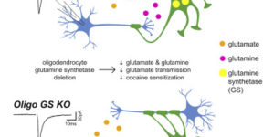
Glutamate has been implicated in a wide range of brain pathologies and is thought to be metabolized via the astrocyte-specific enzyme glutamine synthetase (GS). We show here that oligodendrocytes, the myelinating glia of the central nervous system, also express high levels of GS in caudal regions like the midbrain and the spinal cord. Selective removal of oligodendrocyte GS in mice led to reduced brain glutamate and glutamine levels and impaired glutamatergic synaptic transmission without disrupting myelination. Furthermore, animals lacking oligodendrocyte GS displayed deficits in cocaine-induced locomotor sensitization, a behavior that is dependent on glutamatergic signaling in the midbrain. Thus, oligodendrocytes support glutamatergic transmission through the actions of GS and may represent a therapeutic target for pathological conditions related to brain glutamate dysregulation.

Homeostatic Control of Spontaneous Activity in the Developing Auditory System.

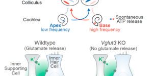
Neurons in the developing auditory system exhibit spontaneous bursts of activity before hearing onset. How this intrinsically generated activity influences development remains uncertain, because few mechanistic studies have been performed in vivo. We show using macroscopic calcium imaging in unanesthetized mice that neurons responsible for processing similar frequencies of sound exhibit highly synchronized activity throughout the auditory system during this critical phase of development. Spontaneous activity normally requires synaptic excitation of spiral ganglion neurons (SGNs). Unexpectedly, tonotopic spontaneous activity was preserved in a mouse model of deafness in which glutamate release from hair cells is abolished. SGNs in these mice exhibited enhanced excitability, enabling direct neuronal excitation by supporting cell-induced potassium transients. These results indicate that homeostatic mechanisms maintain spontaneous activity in the pre-hearing period, with significant implications for both circuit development and therapeutic approaches aimed at treating congenital forms of deafness arising through mutations in key sensory transduction components.

Hair Cell Mechanotransduction Regulates Spontaneous Activity and Spiral Ganglion Subtype Specification in the Auditory System

Type I spiral ganglion neurons (SGNs) transmit sound information from cochlear hair cells to the CNS. Using transcriptome analysis of thousands of single neurons, we demonstrate that murine type I SGNs consist of subclasses that are defined by the expression of subsets of transcription factors, cell adhesion molecules, ion channels, and neurotransmitter receptors. Subtype specification is initiated prior to the onset of hearing during the time period when auditory circuits mature. Gene mutations linked to deafness that disrupt hair cell mechanotransduction or glutamatergic signaling perturb the firing behavior of SGNs prior to hearing onset and disrupt SGN subtype specification. We thus conclude that an intact hair cell mechanotransduction machinery is critical during the pre-hearing period to regulate the firing behavior of SGNs and their segregation into subtypes. Because deafness is frequently caused by defects in hair cells, our findings have significant ramifications for the etiology of hearing loss and its treatment.

Myelin remodeling through experience-dependent oligodendrogenesis in the adult somatosensory cortex.

Oligodendrocyte generation in the adult CNS provides a means to adapt the properties of circuits to changes in life experience. However, little is known about the dynamics of oligodendrocytes and the extent of myelin remodeling in the mature brain. Using longitudinal in vivo two-photon imaging of oligodendrocytes and their progenitors in the mouse cerebral cortex, we show that myelination is an inefficient and extended process, with half of the final complement of oligodendrocytes generated after 4 months of age. Oligodendrocytes that successfully integrated formed new sheaths on unmyelinated and sparsely myelinated axons, and they were extremely stable, gradually changing the pattern of myelination. Sensory enrichment robustly increased oligodendrocyte integration, but did not change the length of existing sheaths. This experience-dependent enhancement of myelination in the mature cortex may accelerate information transfer in these circuits and strengthen the ability of axons to sustain activity by providing additional metabolic support.