Large-scale recording of astrocyte activity.

Astrocytes are highly ramified glial cells found throughout the central nervous system (CNS). They express a variety of neurotransmitter receptors that can induce widespread chemical excitation, placing these cells in an optimal position to exert global effects on brain physiology. However, the activity patterns of only a small fraction of astrocytes have been examined and techniques to manipulate their behavior are limited. As a result, little is known about how astrocytes modulate CNS function on synaptic, microcircuit, or systems levels. Here, we review current and emerging approaches for visualizing and manipulating astrocyte activity in vivo. Deciphering how astrocyte network activity is controlled in different physiological and pathological contexts is crucial for defining their roles in the healthy and diseased CNS.
Early white matter abnormalities, progressive brain pathology and motor deficits in a novel knock-in mouse model of Huntington’s disease.

White matter abnormalities have been reported in premanifest Huntington’s disease (HD) subjects before overt striatal neuronal loss, but whether the white matter changes represent a necessary step towards further pathology and the underlying mechanism of these changes remains unknown. Here, we characterized a novel knock-in mouse model that expresses mouse HD gene homolog (Hdh) with extended CAG repeat- HdhQ250, which was derived from the selective breeding of HdhQ150 mice. HdhQ250 mice manifest an accelerated and robust phenotype compared with its parent line. HdhQ250 mice exhibit progressive motor deficits, reduction in striatal and cortical volume, accumulation of mutant huntingtin aggregation, decreased levels of DARPP32 and BDNF and altered striatal metabolites. The abnormalities detected in this mouse model are reminiscent of several aspects of human HD. In addition, disturbed myelination was evident in postnatal Day 14 HdhQ250 mouse brain, including reduced levels of myelin regulatory factor and myelin basic protein, and decreased numbers of myelinated axons in the corpus callosum. Thinner myelin sheaths, indicated by increased G-ratio of myelin, were also detected in the corpus callosum of adult HdhQ250 mice. Moreover, proliferation of oligodendrocyte precursor cells is altered by mutant huntingtin both in vitro and in vivo. Our data indicate that this model is suitable for understanding comprehensive pathogenesis of HD in white matter and gray matter as well as developing therapeutics for HD.

Human astrocytes develop physiological morphology and remain quiescent in a novel 3D matrix.

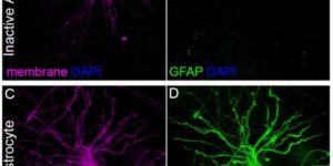
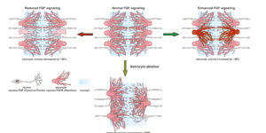
Astrocytes are the most abundant glial cells in the brain and are responsible for diverse functions, from modulating synapse function to regulating the blood-brain barrier. In vivo, these cells exhibit a star-shaped morphology with multiple radial processes that contact synapses and completely surround brain capillaries. In response to trauma or CNS disease, astrocytes become activated, a state associated with profound changes in gene expression, including upregulation of intermediate filament proteins, such as glial fibrillary acidic protein (GFAP). The inability to recapitulate the complex structure of astrocytes and maintain their quiescent state in vitro is a major roadblock to further developments in tissue engineering and regenerative medicine. Here, we characterize astrocyte morphology and activation in various hydrogels to assess the feasibility of developing a matrix that mimics key aspects of the native microenvironment. We show that astrocytes seeded in optimized matrix composed of collagen, hyaluronic acid, and matrigel exhibit a star-shaped morphology with radial processes and do not upregulate GFAP expression, hallmarks of quiescent astrocytes in the brain. In these optimized gels, collagen I provides structural support, HA mimics the brain extracellular matrix, and matrigel provides endothelial cell compatibility and was found to minimize GFAP upregulation. This defined 3D microenvironment for maintaining human astrocytes in vitro provides new opportunities for developing improved models of the blood-brain barrier and studying their response to stress signals.

Calcium dynamics in astrocyte processes during neurovascular coupling.

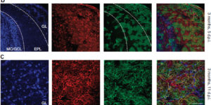
Enhanced neuronal activity in the brain triggers a local increase in blood flow, termed functional hyperemia, via several mechanisms, including calcium (Ca(2+)) signaling in astrocytes. However, recent in vivo studies have questioned the role of astrocytes in functional hyperemia because of the slow and sparse dynamics of their somatic Ca(2+) signals and the absence of glutamate metabotropic receptor 5 in adults. Here, we reexamined their role in neurovascular coupling by selectively expressing a genetically encoded Ca(2+) sensor in astrocytes of the olfactory bulb. We show that in anesthetized mice, the physiological activation of olfactory sensory neuron (OSN) terminals reliably triggers Ca(2+) increases in astrocyte processes but not in somata. These Ca(2+) increases systematically precede the onset of functional hyperemia by 1-2 s, reestablishing astrocytes as potential regulators of neurovascular coupling.

Entrapment via synaptic-like connections between NG2 proteoglycan+ cells and dystrophic axons in the lesion plays a role in regeneration failure after spinal cord injury.

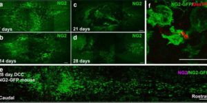
NG2 is purportedly one of the most growth-inhibitory chondroitin sulfate proteoglycans (CSPGs) produced after spinal cord injury. Nonetheless, once the severed axon tips dieback from the lesion core into the penumbra they closely associate with NG2+ cells. We asked if proteoglycans play a role in this tight cell-cell interaction and whether overadhesion upon these cells might participate in regeneration failure in rodents. Studies using varying ratios of CSPGs and adhesion molecules along with chondroitinase ABC, as well as purified adult cord-derived NG2 glia, demonstrate that CSPGs are involved in entrapping neurons. Once dystrophic axons become stabilized upon NG2+ cells, they form synaptic-like connections both in vitro and in vivo. In NG2 knock-out mice, sensory axons in the dorsal columns dieback further than their control counterparts. When axons are double conditioned to enhance their growth potential, some traverse the lesion core and express reduced amounts of synaptic proteins. Our studies suggest that proteoglycan-mediated entrapment upon NG2+ cells is an additional obstacle to CNS axon regeneration.

Spontaneous regeneration of cochlear supporting cells after neonatal ablation ensures hearing in the adult mouse.

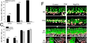
Supporting cells in the cochlea play critical roles in the development, maintenance, and function of sensory hair cells and auditory neurons. Although the loss of hair cells or auditory neurons results in sensorineural hearing loss, the consequence of supporting cell loss on auditory function is largely unknown. In this study, we specifically ablated inner border cells (IBCs) and inner phalangeal cells (IPhCs), the two types of supporting cells surrounding inner hair cells (IHCs) in mice in vivo. We demonstrate that the organ of Corti has the intrinsic capacity to replenish IBCs/IPhCs effectively during early postnatal development. Repopulation depends on the presence of hair cells and cells within the greater epithelial ridge and is independent of cell proliferation. This plastic response in the neonatal cochlea preserves neuronal survival, afferent innervation, and hearing sensitivity in adult mice. In contrast, the capacity for IBC/IPhC regeneration is lost in the mature organ of Corti, and consequently IHC survival and hearing sensitivity are impaired significantly, demonstrating that there is a critical period for the regeneration of cochlear supporting cells. Our findings indicate that the quiescent neonatal organ of Corti can replenish specific supporting cells completely after loss in vivo to guarantee mature hearing function.

A selective thyroid hormone β receptor agonist enhances human and rodent oligodendrocyte differentiation.

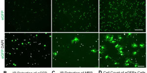
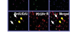
Nerve conduction within the mammalian central nervous system is made efficient by oligodendrocyte-derived myelin. Historically, thyroid hormones have a well described role in regulating oligodendrocyte differentiation and myelination during development; however, it remains unclear which thyroid hormone receptors are required to drive these effects. This is a question with clinical relevance since nonspecific thyroid receptor stimulation can produce deleterious side-effects. Here we report that GC-1, a thyromimetic with selective thyroid receptor β action and a potentially limited side-effect profile, promotes in vitro oligodendrogenesis from both rodent and human oligodendrocyte progenitor cells. In addition, we used in vivo genetic fate tracing of oligodendrocyte progenitor cells via PDGFαR-CreER;Rosa26-eYFP double-transgenic mice to examine the effect of GC-1 on cellular fate and find that treatment with GC-1 during developmental myelination promotes oligodendrogenesis within the corpus callosum, occipital cortex and optic nerve. GC-1 was also observed to enhance the expression of the myelin proteins MBP, CNP and MAG within the same regions. These results indicate that a β receptor selective thyromimetic can enhance oligodendrocyte differentiation in vitro and during developmental myelination in vivo and warrants further study as a therapeutic agent for demyelinating models.

Multiscale optical Ca2+ imaging of tonal organization in mouse auditory cortex.

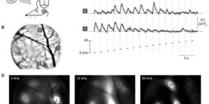
Spatial patterns of functional organization, resolved by microelectrode mapping, comprise a core principle of sensory cortices. In auditory cortex, however, recent two-photon Ca2+ imaging challenges this precept, as the traditional tonotopic arrangement appears weakly organized at the level of individual neurons. To resolve this fundamental ambiguity about the organization of auditory cortex, we developed multiscale optical Ca2+ imaging of unanesthetized GCaMP transgenic mice. Single-neuron activity monitored by two-photon imaging was precisely registered to large-scale cortical maps provided by transcranial widefield imaging. Neurons in the primary field responded well to tones; neighboring neurons were appreciably cotuned, and preferred frequencies adhered tightly to a tonotopic axis. By contrast, nearby secondary-field neurons exhibited heterogeneous tuning. The multiscale imaging approach also readily localized vocalization regions and neurons. Altogether, these findings cohere electrode and two-photon perspectives, resolve new features of auditory cortex, and offer a promising approach generalizable to any cortical area.

Norepinephrine controls astroglial responsiveness to local circuit activity.

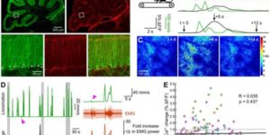
Astrocytes perform crucial supportive functions, including neurotransmitter clearance, ion buffering, and metabolite delivery. They can also influence blood flow and neuronal activity by releasing gliotransmitters in response to intracellular Ca(2+) transients. However, little is known about how astrocytes are engaged during different behaviors in vivo. Here we demonstrate that norepinephrine primes astrocytes to detect changes in cortical network activity. We show in mice that locomotion triggers simultaneous activation of astrocyte networks in multiple brain regions. This global stimulation of astrocytes was inhibited by alpha-adrenoceptor antagonists and abolished by depletion of norepinephrine from the brain. Although astrocytes in visual cortex of awake mice were rarely engaged when neurons were activated by light stimulation alone, pairing norepinephrine release with light stimulation markedly enhanced astrocyte Ca(2+) signaling. Our findings indicate that norepinephrine shifts the gain of astrocyte networks according to behavioral state, enabling astrocytes to respond to local changes in neuronal activity.