NMDA receptor signaling in oligodendrocyte progenitors is not required for oligodendrogenesis and myelination.

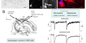
Oligodendrocyte precursor cells (OPCs) express NMDA receptors (NMDARs) and form synapses with glutamatergic neurons throughout the CNS. Although glutamate influences the proliferation and maturation of these progenitors in vitro, the role of NMDAR signaling in oligodendrogenesis and myelination in vivo is not known. Here, we investigated the consequences of genetically deleting the obligatory NMDAR subunit NR1 from OPCs and their oligodendrocyte progeny in the CNS of developing and mature mice. NMDAR-deficient OPCs proliferated normally, achieved appropriate densities in gray and white matter, and differentiated to form major white matter tracts without delay. OPCs also retained their characteristic physiological and morphological properties in the absence of NMDAR signaling and were able to form synapses with glutamatergic axons. However, expression of calcium-permeable AMPA receptors (AMPARs) was enhanced in NMDAR-deficient OPCs. These results suggest that NMDAR signaling is not used to control OPC development but to regulate AMPAR-dependent signaling with surrounding axons, pointing to additional functions for these ubiquitous glial cells.
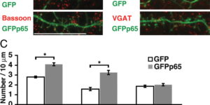
A requirement for nuclear factor-kappaB in developmental and plasticity-associated synaptogenesis.

Structural plasticity of dendritic spines and synapses is a fundamental mechanism governing neuronal circuits and may form an enduring basis for information storage in the brain. We find that the p65 subunit of the nuclear factor-κB (NF-κB) transcription factor, which is required for learning and memory, controls excitatory synapse and dendritic spine formation and morphology in murine hippocampal neurons. Endogenous NF-κB activity is elevated by excitatory transmission during periods of rapid spine and synapse development. During in vitro synaptogenesis, NF-κB enhances dendritic spine and excitatory synapse density and loss of endogenous p65 decreases spine density and spine head volume. Cell-autonomous function of NF-κB within the postsynaptic neuron is sufficient to regulate the formation of both presynaptic and postsynaptic elements. During synapse development in vivo, loss of NF-κB similarly reduces spine density and also diminishes the amplitude of synaptic responses. In contrast, after developmental synaptogenesis has plateaued, endogenous NF-κB activity is low and p65 deficiency no longer attenuates basal spine density. Instead, NF-κB in mature neurons is activated by stimuli that induce demand for new synapses, including estrogen and short-term bicuculline, and is essential for upregulating spine density in response to these stimuli. p65 is enriched in dendritic spines making local protein-protein interactions possible; however, the effects of NF-κB on spine density require transcription and the NF-κB-dependent regulation of PSD-95, a critical postsynaptic component. Collectively, our data define a distinct role for NF-κB in imparting transcriptional regulation required for the induction of changes to, but not maintenance of, excitatory synapse and spine density.

NG2+ CNS glial progenitors remain committed to the oligodendrocyte lineage in postnatal life and following neurodegeneration.

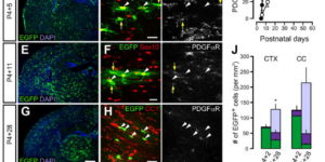
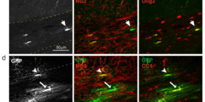
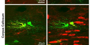
The mammalian CNS contains a ubiquitous population of glial progenitors known as NG2+ cells that have the ability to develop into oligodendrocytes and undergo dramatic changes in response to injury and demyelination. Although it has been reported that NG2+ cells are multipotent, their fate in health and disease remains controversial. Here, we generated PDGFαR-CreER transgenic mice and followed their fate in vivo in the developing and adult CNS. These studies revealed that NG2+ cells in the postnatal CNS generate myelinating oligodendrocytes, but not astrocytes or neurons. In regions of neurodegeneration in the spinal cord of ALS mice, NG2+ cells exhibited enhanced proliferation and accelerated differentiation into oligodendrocytes but remained committed to the oligodendrocyte lineage. These results indicate that NG2+ cells in the normal CNS are oligodendrocyte precursors with restricted lineage potential and that cell loss and gliosis are not sufficient to alter the lineage potential of these progenitors.

Calcium action potentials in hair cells pattern auditory neuron activity before hearing onset.

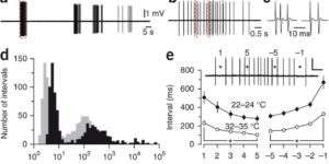
We found rat central auditory neurons to fire action potentials in a precise sequence of mini-bursts before the age of hearing onset. This stereotyped pattern was initiated by hair cells in the cochlea, which trigger brief bursts of action potentials in auditory neurons each time they fire a Ca2+ spike. By generating theta-like activity, hair cells may limit the influence of synaptic depression in developing auditory circuits and promote consolidation of synapses.

ATP-induced morphological changes in supporting cells of the developing cochlea.

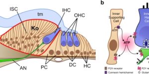
The developing cochlea of mammals contains a large group of columnar-shaped cells, which together form a structure known as Kölliker’s organ. Prior to the onset of hearing, these inner supporting cells periodically release adenosine 5′-triphosphate (ATP), which activates purinergic receptors in surrounding supporting cells, inner hair cells and the dendrites of primary auditory neurons. Recent studies indicate that purinergic signaling between inner supporting cells and inner hair cells initiates bursts of action potentials in auditory nerve fibers before the onset of hearing. ATP also induces prominent effects in inner supporting cells, including an increase in membrane conductance, a rise in intracellular Ca(2+), and dramatic changes in cell shape, although the importance of ATP signaling in non-sensory cells of the developing cochlea remains unknown. Here, we review current knowledge pertaining to purinergic signaling in supporting cells of Kölliker’s organ and focus on the mechanisms by which ATP induces changes in their morphology. We show that these changes in cell shape are preceded by increases in cytoplasmic Ca(2+), and provide new evidence indicating that elevation of intracellular Ca(2+) and IP(3) are sufficient to initiate shape changes. In addition, we discuss the possibility that these ATP-mediated morphological changes reflect crenation following the activation of Ca(2+)-activated Cl(-) channels, and speculate about the possible functions of these changes in cell morphology for maturation of the cochlea.

Zones of enhanced glutamate release from climbing fibers in the mammalian cerebellum.

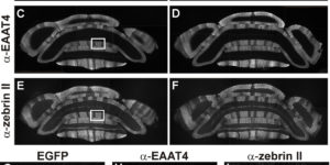
Purkinje cells in the mammalian cerebellum are remarkably homogeneous in shape and orientation, yet they exhibit regional differences in gene expression. Purkinje cells that express high levels of zebrin II (aldolase C) and the glutamate transporter EAAT4 cluster in parasagittal zones that receive input from distinct groups of climbing fibers (CFs); however, the physiological properties of CFs that target these molecularly distinct Purkinje cells have not been determined. Here we report that CFs that innervate Purkinje cells in zebrin II-immunoreactive (Z(+)) zones release more glutamate per action potential than CFs in Z(-) zones. CF terminals in Z(+) zones had larger pools of release-ready vesicles, exhibited enhanced multivesicular release, and produced larger synaptic glutamate transients. As a result, CF-mediated EPSCs in Purkinje cells decayed more slowly in Z(+) zones, which triggered longer-duration complex spikes containing a greater number of spikelets. The differences in the duration of CF EPSCs between Z(+) and Z(-) zones persisted in EAAT4 knock-out mice, indicating that EAAT4 is not required for maintaining this aspect of CF function. These results indicate that the organization of the cerebellum into discrete longitudinal zones is defined not only by molecular phenotype of Purkinje cells within zones, but also by the physiological properties of CFs that project to these distinct regions. The enhanced release of glutamate from CFs in Z(+) zones may alter the threshold for synaptic plasticity and prolong inhibition of cerebellar output neurons in deep cerebellar nuclei.

Neuron-glia synapses in the brain

The ability to investigate the electrophysiological properties of individual cells in acute brain tissue led to the discovery that many glial cells have the capacity to respond rapidly to neuronal activity. In particular, a distinct class of neuroglial cells known as NG2 cells, which exhibit many of the properties that have been described for glial subtypes such as complex cells, polydendrocytes, synantocytes and GluR cells, express ionotropic receptors for glutamate and GABA. In both gray and white matter, NG2 cells form direct synaptic junctions with axons, which enable transient activation of these receptors. Electrophysiological analyses have shown that these neuron-glia synapses exhibit all the hallmarks of ‘classical’ neuron-neuron synapses, including rapid activation, quantized responses, facilitation and depression, and presynaptic inhibition. Electron microscopy indicates that axons form morphologically distinct junctions at discrete sites along processes of NG2 cells, suggesting that NG2 cells are an overt target of axonal projections. AMPA receptors expressed by NG2 cells exhibit varying degrees of Ca(2+) permeability, depending on the brain region and stage of development, and in white matter NG2 cells have also been shown to express functional NMDA receptors. Ca(2+) influx through AMPA receptors following repetitive stimulation can trigger long term potentiation of synaptic currents in NG2 cells. The expression of receptors with significant Ca(2+) permeability may increase the susceptibility of NG2 cells to excitotoxic injury. Future studies using transgenic mice in which expression of receptors can be manipulated selectively in NG2 cells have to define the functions of this enigmatic neuron-glia signaling in the normal and diseased CNS.

Excitability and synaptic communication within the oligodendrocyte lineage

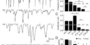
The mammalian CNS contains an abundant, widely distributed population of glial cells that serve as oligodendrocyte progenitors. It has been reported that these NG2-immunoreactive cells (NG2(+) cells) form synapses and generate action potentials, suggesting that neural-evoked excitation of these progenitors may regulate oligodendrogenesis. However, recent studies also suggest that NG2(+) cells are comprised of functionally distinct groups that differ in their ability to respond to neuronal activity, undergo differentiation, and experience injury following ischemia. To better define the physiological properties of NG2(+) cells, we used transgenic mice that allowed an unbiased sampling of this population and unambiguous identification of cells in discrete states of differentiation. Using acute brain slices prepared from developing and mature mice, we found that NG2(+) cells in diverse brain regions share a core set of physiological properties, including expression of voltage-gated Na(+) (NaV) channels and ionotropic glutamate receptors, and formation of synapses with glutamatergic neurons. Although small amplitude Na(+) spikes could be elicited in some NG2(+) cells during the first postnatal week, they were not capable of generating action potentials. Transition of these progenitors to the premyelinating stage was accompanied by the rapid removal of synaptic input, as well as downregulation of AMPA and NMDA receptors and NaV channels. Thus, prior reports of physiological heterogeneity among NG2(+) cells may reflect analysis of cells in later stages of maturation. These results suggest that NG2(+) cells are uniquely positioned within the oligodendrocyte lineage to monitor the firing patterns of surrounding neurons.

Developmental regulation of spontaneous activity in the Mammalian cochlea.

Neurons in the developing auditory system fire bursts of action potentials before the onset of hearing. This spontaneousactivity promotes the survival and maturation of auditory neurons and the refinement of synaptic connections in auditory nuclei; however, the mechanisms responsible for initiating this activity remain uncertain. Previous studies indicate that inner supporting cells (ISCs) in the developing cochlea periodically release ATP, which depolarizes inner hair cells (IHCs), leading to bursts of action potentials in postsynaptic spiral ganglion neurons (SGNs). To determine when purinergic signaling appears in the developing cochlea and whether it is responsible for initiating auditory neuron activity throughout the prehearing period, we examined spontaneousactivity from ISCs, IHCs, and SGNs in cochleae acutely isolated from rats during the first three postnatal weeks. We found that ATP was released from ISCs within the cochlea from birth until the onset of hearing, which led to periodic inward currents, Ca(2+) transients, and morphological changes in these supporting cells. This spontaneous release of ATP also depolarized IHCs and triggered bursts of action potentials in SGNs for most of the postnatal prehearing period, beginning a few days after birth as IHCs became responsive to ATP, until the onset of hearing when ATP was no longer released from ISCs. When IHCs were not subject to purinergic excitation, SGNs exhibited little or no activity. These results suggest that supporting cells in the cochlea provide the primary excitatory stimulus responsible for initiating bursts of action potentials in auditory nerve fibers before the onset of hearing.

Photon capture and signalling by melanopsin retinal ganglion cells

A subset of retinal ganglion cells has recently been discovered to be intrinsically photosensitive, with melanopsin as the pigment. These cells project primarily to brain centres for non-image-forming visual functions such as the pupillary light reflex and circadian photoentrainment. How well they signal intrinsic light absorption to drive behaviour remains unclear. Here we report fundamental parameters governing their intrinsic light responses and associated spike generation. The membrane density of melanopsin is 10(4)-fold lower than that of rod and cone pigments, resulting in a very low photon catch and a phototransducing role only in relatively bright light. Nonetheless, each captured photon elicits a large and extraordinarily prolonged response, with a unique shape among known photoreceptors. Notably, like rods, these cells are capable of signalling single-photon absorption. A flash causing a few hundred isomerized melanopsin molecules in a retina is sufficient for reaching threshold for the pupillary light reflex.