Ncm-D-aspartate: a novel caged D-aspartate suitable for activation of glutamate transporters and N-methyl-D-aspartate (NMDA) receptors in brain tissue

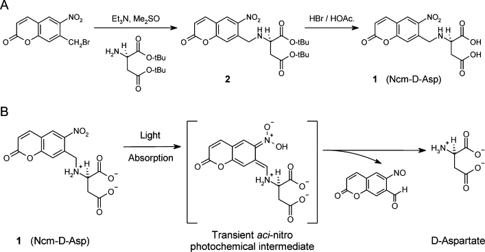
The D-isomer of aspartate is both a substrate for glutamate transporters and an agonist of N-methyl-D-aspartate (NMDA) receptors. To monitor the behavior of these receptors and transporters in intact tissue we synthesized a new photo-labile analogue of D-aspartate, N-[(6-nitrocoumarin-7-yl)methyl]-D-aspartic acid (Ncm-D-aspartate). This compound was photolyzed rapidly (t(1/2)=0.11 micros) by UV light with a quantum efficiency of 0.041 at pH 7.4. In acute hippocampal slices, photolysis of Ncm-D-aspartate by brief (1 ms) exposure to UV light elicited rapidly activating inward currents in astrocytes that were sensitive to inhibition by the glutamate transporter antagonist DL-threo-beta-benzyloxyaspartic acid (TBOA). Neither Ncm-D-aspartate nor the photo-released caging group exhibited agonist or antagonist activity at glutamate transporters, and Ncm-D-aspartate did not induce transporter currents prior to photolysis. Glutamate transporter currents were also elicited in cerebellar Purkinje cells in response to photolysis of Ncm-D-aspartate. Photo-release of D-aspartate from Ncm-D-aspartate did not induce alpha-amino-3-hydroxy-5-methyl-4-isoxazole propionic acid (AMPA)/kainate receptor or metabotropic glutamate receptor (mGluR) currents, but triggered robust NMDA receptor currents in neurons; Ncm-D-aspartate and the photolzyed caging group were similarly inert at NMDA receptors. These results indicate that Ncm-D-aspartate can be used to study NMDA receptors at excitatory synapses and interactions between transporters and receptors in brain tissue.
Climbing fiber innervation of NG2-expressing glia in the mammalian cerebellum

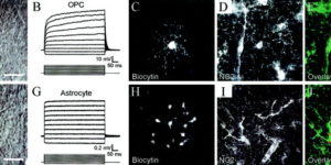
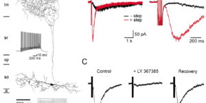
The molecular layer of the cerebellar cortex is populated by glial progenitors that express ionotropic glutamate receptors and extend numerous processes among Purkinje cell dendrites. Here, we show that release of glutamate from climbing fiber (CF) axons produces AMPA receptor currents with rapid kinetics in these NG2-immunoreactive glial cells (NG2+ cells) in cerebellar slices. NG2+ cells may receive up to 70 discrete inputs from one CF and, unlike mature Purkinje cells, are often innervated by multiple CFs. Paired Purkinje cell-NG2+ cell recordings show that one CF can innervate both cell types. CF boutons make direct synaptic junctions with NG2+ cell processes, indicating that this rapid neuron-glia signaling occurs at discrete sites rather than through ectopic release at CF-Purkinje cell synapses. This robust activation of Ca2+-permeable AMPA receptors in NG2+ cells expands the influence of the olivocerebellar projection to this abundant class of glial progenitors.

Synthesis and characterization of 4-methoxy-7-nitroindolinyl-D-aspartate, a caged compound for selective activation of glutamate transporters and N-methyl-D-aspartate receptors in brain tissue.

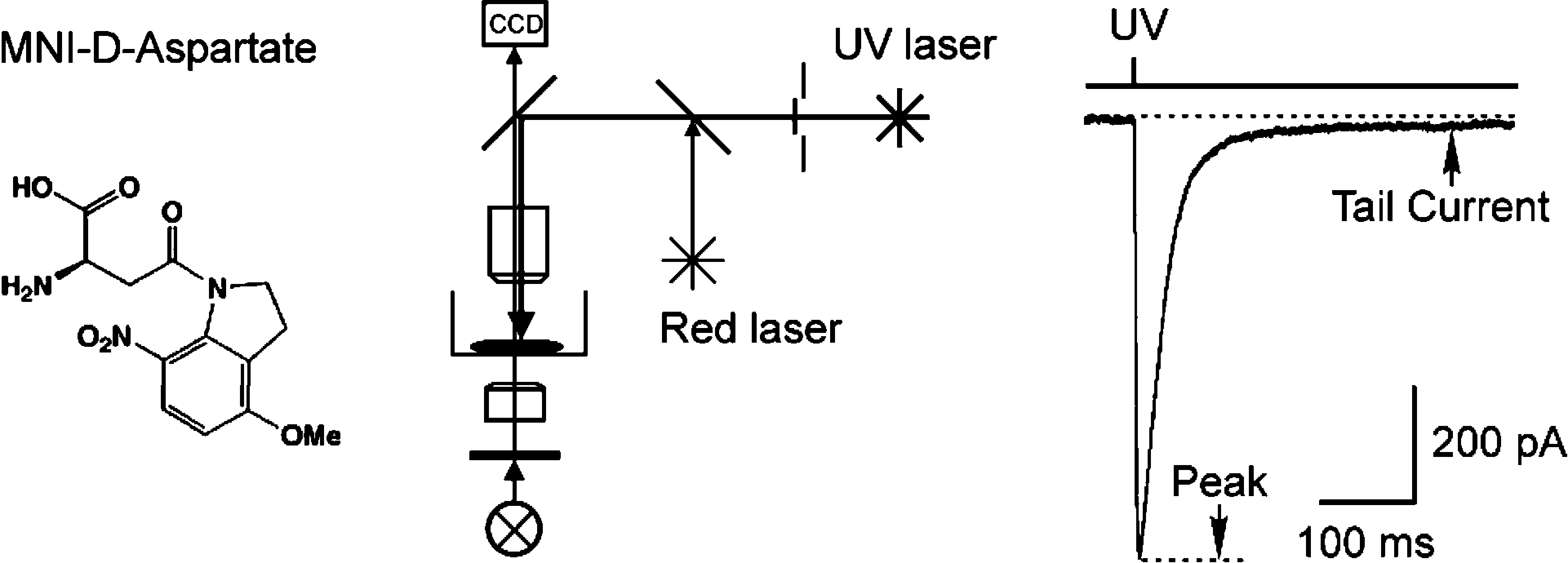
The D-isomer of aspartate is efficiently transported by high-affinity Na(+)/K(+)-dependent glutamate transporters and is an effective ligand of N-methyl-d-aspartate (NMDA) receptors. To facilitate analysis of the regulation of these proteins in their native membranes, we synthesized a photolabile analogue of D-aspartate, 4-methoxy-7-nitroindolinyl-D-aspartate (MNI-D-aspartate). This compound was photolyzed with a quantum efficiency of 0.09 at pH 7.4. Photorelease of d-aspartate in acute hippocampal slices through brief (1 ms) UV laser illumination of MNI-d-aspartate triggered rapidly activating currents in astrocytes that were inhibited by the glutamate transporter antagonist DL-threo-beta-benzyloxyaspartic acid (TBOA), indicating that they resulted from electrogenic uptake of D-aspartate. These transporter currents exhibited a distinct tail component that was approximately 2% of the peak current, which may result from the release of K(+) into the extracellular space during counter transport. MNI-D-aspartate was neither an agonist nor an antagonist of glutamate transporters at concentrations up to 500 muM and was stable in aqueous solution for several days. Glutamate transporter currents were also elicited in Bergmann glial cells and Purkinje neurons of the cerebellum in response to photolysis of MNI-D-aspartate, indicating that this compound can be used for monitoring the occupancy and regulation of glutamate transporters in different brain regions. Photorelease of D-aspartate did not activate alpha-amino-3-hydroxy-5-methyl-4-isoxazolepropionic acid (AMPA)/kainate receptors or metabotropic glutamate receptors (mGluRs) in neurons, but resulted in the selective, but transient, activation of NMDA receptors in hippocampal pyramidal neurons; MNI-D-aspartate was not an antagonist of NMDA receptors. These results indicate that MNI-D-aspartate also may be useful for studying the regulation of NMDA receptors at excitatory synapses.

Beta-lactam antibiotics offer neuroprotection by increasing glutamate transporter expression.

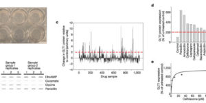
Glutamate is the principal excitatory neurotransmitter in the nervous system. Inactivation of synaptic glutamate is handled by the glutamate transporter GLT1 (also known as EAAT2; refs 1, 2), the physiologically dominant astroglial protein. In spite of its critical importance in normal and abnormal synaptic activity, no practical pharmaceutical can positively modulate this protein. Animal studies show that the protein is important for normal excitatory synaptic transmission, while its dysfunction is implicated in acute and chronic neurological disorders, including amyotrophic lateral sclerosis (ALS), stroke, brain tumours and epilepsy. Using a blinded screen of 1,040 FDA-approved drugs and nutritionals, we discovered that many beta-lactam antibiotics are potent stimulators of GLT1 expression. Furthermore, this action appears to be mediated through increased transcription of the GLT1 gene. beta-Lactams and various semi-synthetic derivatives are potent antibiotics that act to inhibit bacterial synthetic pathways. When delivered to animals, the beta-lactam ceftriaxone increased both brain expression of GLT1 and its biochemical and functional activity. Glutamate transporters are important in preventing glutamate neurotoxicity. Ceftriaxone was neuroprotective in vitro when used in models of ischaemic injury and motor neuron degeneration, both based in part on glutamate toxicity. When used in an animal model of the fatal disease ALS, the drug delayed loss of neurons and muscle strength, and increased mouse survival. Thus these studies provide a class of potential neurotherapeutics that act to modulate the expression of glutamate neurotransmitter transporters via gene activation.

Synaptic signaling between neurons and glia.

Rapid signaling between vertebrate neurons occurs primarily at synapses, intercellular junctions where quantal release of neurotransmitter triggers rapid changes in membrane conductance through activation of ionotropic receptors. Glial cells express many of these same ionotropic receptors, yet little is known about how receptors in glial cells become activated in situ. Because synapses were thought to be the sole provenance of neurons, it has been assumed that these receptors must be activated following diffusion of transmitter out of the synaptic cleft, or through nonsynaptic mechanisms such as transporter reversal. Two recent reports show that a ubiquitous class of progenitors that express the proteoglycan NG2 (NG2 cells) engage in rapid signaling with glutamatergic and gamma-aminobutyric acid (GABA)ergic neurons through direct neuron-glia synapses. Quantal release of transmitter from neurons at these sites triggers rapid activation of aminomethylisoxazole propionic acid (AMPA) or GABA(A) receptors in NG2 cells. These currents exhibit properties consistent with direct rather than spillover-mediated transmission, and electron micrographic analyses indicate that nerve terminals containing clusters of synaptic vesicles form discrete junctions with NG2 cell processes. Although activation of AMPA or GABA(A) receptors depolarize NG2 cells, these receptors are more likely to serve as routes for ion flux rather than as current sources for depolarization, because the amplitudes of the synaptic transients are small and the resting membrane potential of NG2 cells is highly negative. The ability of both glutamate and GABA to influence the morphology, physiology, and development of NG2 cells in vitro suggests that this rapid form of signaling may play important roles in adapting the behavior of these cells to the needs of surrounding neurons in vivo.

Glutamate transporters bring competition to the synapse

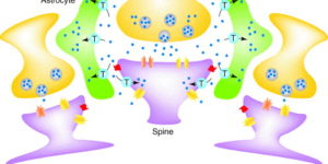
Glutamate transporters (GluTs) prevent the accumulation of glutamate and influence the occupancy of receptors at synapses. The ability of extrasynaptic NMDA receptors and metabotropic glutamate receptors to participate in signaling is tightly regulated by GluT activity. Astrocytes express the highest density of GluTs and dominate clearance away from these receptors; synapses that are not associated with astrocyte processes experience greater mGluR activation and can be exposed to glutamate released at adjacent synapses. Although less abundant, neuronal transporters residing in the postsynaptic membrane can also shield receptors from the glutamate that is released. The diversity in synaptic morphology suggests a correspondingly rich diversity of GluT function in excitatory transmission.

Astrocyte glutamate transporters regulate metabotropic glutamate receptor-mediated excitation of hippocampal interneurons.

Clearance of extracellular glutamate is essential for limiting the activity of metabotropic glutamate receptors (mGluRs) at excitatory synapses; however, the relative contribution of transporters found in neuronal and glial membranes to this uptake is poorly understood. Hippocampalinterneurons located at the oriens-alveus border express mGluR1alpha, a metabotropic glutamate receptor that regulates excitability and synaptic plasticity. To determine which glutamate transporters are essential for removing glutamate at these excitatory synapses, we recorded mGluR1-mediated EPSCs from oriens-lacunosum moleculare (O-LM) interneurons in acute hippocampal slices. Stimulation in stratum oriens reliably elicited a slow mGluR1-mediated current in O-LM interneurons if they were briefly depolarized to allow Ca2+ entry before stimulation. Selective inhibition of GLT-1 [for glutamate transporter; EAAT2 (for excitatory amino acid transporter)] with dihydrokainate increased the amplitude of these responses approximately threefold, indicating that these transporters compete with mGluRs for synaptically released glutamate. However, inhibition of all glutamate transporters with TBOA (DL-threo-b-benzyloxyaspartic acid) increased mGluR1 EPSCs >15-fold, indicating that additional transporters also shape activation of these receptors. To identify these transporters, we examined mGluR1 EPSCs in mice lacking GLAST (for glutamate-aspartate transporter; EAAT1) or EAAC1 (for excitatory amino acid carrier; EAAT3). A comparison of responses recorded from wild-type and transporter knock-out mice revealed that the astroglial glutamate transporters GLT-1 and GLAST, but not the neuronal transporter EAAC1, restrict activation of mGluRs in O-LM interneurons. Transporter-dependent potentiation of mGluR1 EPSCs led to a dramatic increase in interneuron firing and enhanced inhibition of CA1 pyramidal neurons, suggesting that acute or prolonged disruption of transporter activity could lead to changes in network activity as a result of enhanced interneuron excitability.

Climbing fiber activation of EAAT4 transporters and kainate receptors in cerebellar Purkinje cells

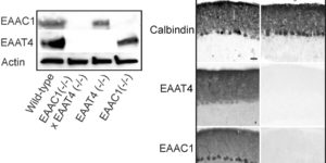
Cerebellar Purkinje cells (PCs) express two glutamate transporters, EAAC1 (EAAT3) and EAAT4; however, their relative contribution to the uptake of glutamate at synapses is not known. We found that glutamate transporter currents recorded at climbing fiber (CF)-PC synapses are absent in mice lacking EAAT4 but unchanged in mice lacking EAAC1, indicating that EAAT4 is preferentially involved in clearing glutamate from CF synapses. However, comparison of CF synaptic currents between wild-type and transporter knock-out mice revealed that ionotropic glutamate receptors are responsible for >40% of the current previously attributed to transporters, indicating that PCs remove <10% of the glutamate released by the CF. The receptors responsible for the nontransporter component accounted for 5% of the CF EPSC, had a slower time course and lower occupancy than AMPA receptors at CF synapses, and exhibited pharmacological properties consistent with kainate receptors. In GluR5 knock-out mice, this current was dramatically reduced, indicating that CF excitation of PCs involves two distinct classes of ionotropic glutamate receptors, AMPA receptors and GluR5-containing kainate receptors.

Synaptic signaling between GABAergic interneurons and oligodendrocyte precursor cells in the hippocampus

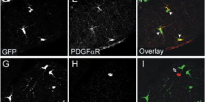
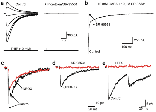
Oligodendrocyte precursor cells (OPCs) express receptors for many neurotransmitters, but the mechanisms responsible for their activation are poorly understood. We have found that quantal release of GABA from interneurons elicits GABA(A) receptor currents with rapid rise times in hippocampal OPCs. These currents did not exhibit properties of spillover transmission or release by transporters, and immunofluorescence and electron microscopy suggest that interneuronal terminals are in direct contact with OPCs, indicating that these GABA currents are generated at direct interneuron-OPC synapses. The reversal potential of OPC GABA(A) currents was -43 mV, and interneuronal firing was correlated with transient depolarizations induced by GABA(A) receptors; however, GABA application induced a transient inhibition of currents mediated by AMPA receptors in OPCs. These results indicate that OPCs are a direct target of interneuronal collaterals and that the GABA-induced Cl(-) flux generated by these events may influence oligodendrocyte development by regulating the efficacy of glutamatergic signaling in OPCs.