Glutamate transporters bring competition to the synapse

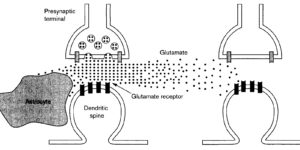
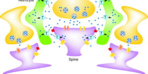
Glutamate transporters (GluTs) prevent the accumulation of glutamate and influence the occupancy of receptors at synapses. The ability of extrasynaptic NMDA receptors and metabotropic glutamate receptors to participate in signaling is tightly regulated by GluT activity. Astrocytes express the highest density of GluTs and dominate clearance away from these receptors; synapses that are not associated with astrocyte processes experience greater mGluR activation and can be exposed to glutamate released at adjacent synapses. Although less abundant, neuronal transporters residing in the postsynaptic membrane can also shield receptors from the glutamate that is released. The diversity in synaptic morphology suggests a correspondingly rich diversity of GluT function in excitatory transmission.
Astrocyte glutamate transporters regulate metabotropic glutamate receptor-mediated excitation of hippocampal interneurons.

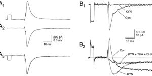
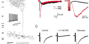
Clearance of extracellular glutamate is essential for limiting the activity of metabotropic glutamate receptors (mGluRs) at excitatory synapses; however, the relative contribution of transporters found in neuronal and glial membranes to this uptake is poorly understood. Hippocampalinterneurons located at the oriens-alveus border express mGluR1alpha, a metabotropic glutamate receptor that regulates excitability and synaptic plasticity. To determine which glutamate transporters are essential for removing glutamate at these excitatory synapses, we recorded mGluR1-mediated EPSCs from oriens-lacunosum moleculare (O-LM) interneurons in acute hippocampal slices. Stimulation in stratum oriens reliably elicited a slow mGluR1-mediated current in O-LM interneurons if they were briefly depolarized to allow Ca2+ entry before stimulation. Selective inhibition of GLT-1 [for glutamate transporter; EAAT2 (for excitatory amino acid transporter)] with dihydrokainate increased the amplitude of these responses approximately threefold, indicating that these transporters compete with mGluRs for synaptically released glutamate. However, inhibition of all glutamate transporters with TBOA (DL-threo-b-benzyloxyaspartic acid) increased mGluR1 EPSCs >15-fold, indicating that additional transporters also shape activation of these receptors. To identify these transporters, we examined mGluR1 EPSCs in mice lacking GLAST (for glutamate-aspartate transporter; EAAT1) or EAAC1 (for excitatory amino acid carrier; EAAT3). A comparison of responses recorded from wild-type and transporter knock-out mice revealed that the astroglial glutamate transporters GLT-1 and GLAST, but not the neuronal transporter EAAC1, restrict activation of mGluRs in O-LM interneurons. Transporter-dependent potentiation of mGluR1 EPSCs led to a dramatic increase in interneuron firing and enhanced inhibition of CA1 pyramidal neurons, suggesting that acute or prolonged disruption of transporter activity could lead to changes in network activity as a result of enhanced interneuron excitability.

Climbing fiber activation of EAAT4 transporters and kainate receptors in cerebellar Purkinje cells

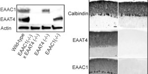
Cerebellar Purkinje cells (PCs) express two glutamate transporters, EAAC1 (EAAT3) and EAAT4; however, their relative contribution to the uptake of glutamate at synapses is not known. We found that glutamate transporter currents recorded at climbing fiber (CF)-PC synapses are absent in mice lacking EAAT4 but unchanged in mice lacking EAAC1, indicating that EAAT4 is preferentially involved in clearing glutamate from CF synapses. However, comparison of CF synaptic currents between wild-type and transporter knock-out mice revealed that ionotropic glutamate receptors are responsible for >40% of the current previously attributed to transporters, indicating that PCs remove <10% of the glutamate released by the CF. The receptors responsible for the nontransporter component accounted for 5% of the CF EPSC, had a slower time course and lower occupancy than AMPA receptors at CF synapses, and exhibited pharmacological properties consistent with kainate receptors. In GluR5 knock-out mice, this current was dramatically reduced, indicating that CF excitation of PCs involves two distinct classes of ionotropic glutamate receptors, AMPA receptors and GluR5-containing kainate receptors.

Synaptic signaling between GABAergic interneurons and oligodendrocyte precursor cells in the hippocampus

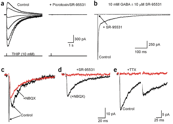
Oligodendrocyte precursor cells (OPCs) express receptors for many neurotransmitters, but the mechanisms responsible for their activation are poorly understood. We have found that quantal release of GABA from interneurons elicits GABA(A) receptor currents with rapid rise times in hippocampal OPCs. These currents did not exhibit properties of spillover transmission or release by transporters, and immunofluorescence and electron microscopy suggest that interneuronal terminals are in direct contact with OPCs, indicating that these GABA currents are generated at direct interneuron-OPC synapses. The reversal potential of OPC GABA(A) currents was -43 mV, and interneuronal firing was correlated with transient depolarizations induced by GABA(A) receptors; however, GABA application induced a transient inhibition of currents mediated by AMPA receptors in OPCs. These results indicate that OPCs are a direct target of interneuronal collaterals and that the GABA-induced Cl(-) flux generated by these events may influence oligodendrocyte development by regulating the efficacy of glutamatergic signaling in OPCs.

Comparison of coupled and uncoupled currents during glutamate uptake by GLT-1 transporters

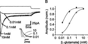
The transport of glutamate across the plasma membrane is coupled to the movement of cations (Na+, K+, and H+) that are necessary for glutamate uptake and transporter cycling as well as anions that are uncoupled from the flux of glutamate. Although the relationship between these coupled (stoichiometric) and uncoupled (anion) transporter currents is poorly understood, transporter-associated anion currents often are used to monitor transporter activity. To define the kinetic relationship between these two components, we have recorded transporter currents associated with stoichiometric and anion charge movements occurring in response to the rapid application of l-glutamate to outside-out patches from human embryonic kidney cells expressing GLT-1 transporters. Transporter-associated anion currents were approximately twice as slow to rise and decay as stoichiometric transport currents, but the presence of permeant anions did not slow transporter cycling. A kinetic model for GLT-1 was developed to simulate the behavior of both components of the transporter current and to estimate the capture efficiency of GLT-1. In this model the K+ counter-transport step was defined as rate-limiting, consistent with the slowing of transporter cycling after the substitution of internal K+ with Cs+ or Na+. The model predicts that in physiological conditions approximately 35% of GLT-1 transporters function as buffers, releasing glutamate back into the extracellular space after binding.

Physiological characteristics of NG2-expressing glial cells.

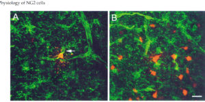
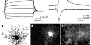
Antibodies against the chondroitin sulfate proteoglycan NG2 label a subpopulation of glial cells within the CNS, which have a small cell body and thin radiating processes. Physiological recordings from these small cells in acute brain slices have revealed that they possess unique properties, suggesting that they may comprise a class of glial cells distinct from astrocytes, oligodendrocytes, or microglia. NG2-expressing glial cells (abbreviated as “NG2 cells” here) have a moderate input resistance and are not dye- or tracer-coupled to adjacent cells. They express voltage-gated Na+, K+ and Ca2+ conductances, though they do not exhibit regenerative Na+ or Ca2+ action potentials due to the much larger K+ conductances present. In addition to voltage-gated conductances, they express receptors for various neurotransmitters. In the hippocampus, AMPA and GABAA receptors on these cells are activated by release of transmitter from neurons at defined synaptic junctions that are formed with CA3 pyramidal neurons and GABAergic interneurons. These rapid forms of neuron-glial communication may regulate the proliferation rate of NG2 cells or their development into mature oligodendrocytes. These depolarizing inputs may also trigger the release of neuroactive substances from NG2 cells, providing feedback regulation of signaling at neuronal synapses. Although the presence of Ca2+ permeable AMPA receptors provides a pathway to link neuronal activity to Ca2+ dependent processes within the NG2 cells, these receptors also put these cells at risk for glutamate-associated excitotoxicity. This vulnerability to the sustained elevation of glutamate may underlie ischemic induced damage to white matter tracts and contribute to cerebral palsy in premature infants.

Glutamatergic synapses on oligodendrocyte precursor cells in the hippocampus.

Fast excitatory neurotransmission in the central nervous system occurs at specialized synaptic junctions between neurons, where a high concentration of glutamate directly activates receptor channels. Low-affinity AMPA (alpha-amino-3-hydroxy-5-methyl isoxazole propionic acid) and kainate glutamate receptors are also expressed by some glial cells, including oligodendrocyte precursor cells (OPCs). However, the conditions that result in activation of glutamate receptors on these non-neuronal cells are not known. Here we report that stimulation of excitatory axons in the hippocampus elicits inward currents in OPCs that are mediated by AMPA receptors. The quantal nature of these responses and their rapid kinetics indicate that they are produced by the exocytosis of vesicles filled with glutamate directly opposite these receptors. Some of these AMPA receptors are permeable to calcium ions, providing a link between axonal activity and internal calcium levels in OPCs. Electron microscopic analysis revealed that vesicle-filled axon terminals make synaptic junctions with the processes of OPCs in both the young and adult hippocampus. These results demonstrate the existence of a rapid signalling pathway from pyramidal neurons to OPCs in the mammalian hippocampus that is mediated by excitatory, glutamatergic synapses.

Clearance of glutamate inside the synapse and beyond

The heated debate over the level of postsynaptic receptor occupancy by transmitter has not been extinguished – indeed, new evidence is fanning the flames. Recent experiments using two-photon microscopy suggest that the concentration of glutamate in the synaptic cleft does not attain levels previously suggested. In contrast, recordings from glial cells and studies of extrasynaptic receptor activation indicate that significant quantities of glutamate escape from the cleft following exocytosis. Determining the amount of glutamate efflux from the synaptic cleft and the distance it diffuses is critical to issues of synaptic specificity and the induction of synaptic plasticity.

Glial contribution to glutamate uptake at Schaffer collateral-commissural synapses in the hippocampus

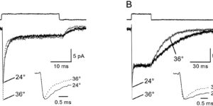
Astrocytes in the hippocampus express high-affinity glutamate transporters that are important for lowering the concentration of extracellular glutamate after release at excitatory synapses. These transporters exhibit a permeability to chaotropic anions that is associated with transport, allowing their activity to be monitored in cell-fee patches when highly permeant anions are present. Astrocyte glutamate transporters are highly temperature sensitive, because L-glutamate-activated, anion-potentiated transporter currents in outside-out patches from these cells exhibited larger amplitudes and faster kinetics at 36 degreesC than at 24 degreesC. The cycling rate of these transporters was estimated by using paired applications of either L-glutamate or D-aspartate to measure the time necessary for the peak of the transporter current to recover from the steady-state level. Transporter currents in patches recovered with a time constant of 11.6 msec at 36 degreesC, suggesting that either the turnover rate of native transporters is much faster than previously reported for expressed EAAT2 transporters or the efficiency of these transporters is very low. Synaptically activated transporter currents persisted in astrocytes at physiological temperatures, although no evidence of these currents was found in CA1 pyramidal neurons in response to afferent stimulation. L-glutamate-gated transporter currents were also not detected in outside-out patches from pyramidal neurons. These results are consistent with the hypothesis that astrocyte transporters are responsible for taking up the majority of glutamate released at Schaffer collateral-commissural synapses in the hippocampus.Glial contribution to glutamate uptake at Schaffer collateral-commissural synapses in the hippocampus

Glutamate Release Monitored with Astrocyte Transporter Currents during LTP

Long-term potentiation (LTP) of synaptic transmission in the CA1 region of the hippocampus is thought to result from either increased transmitter release, heightened postsynaptic sensitivity, or a combination of the two. We have measured evoked glutamate release from Schaffer collateral/commissural fiber terminals in CA1 by recording synaptically activated glutamate transporter currents in hippocampal astrocytes located in stratum radiatum. Although several manipulations of release probability caused parallel changes in extracellular field potentials and synaptically activated transporter current amplitudes, induction of LTP failed to alter transporter-mediated responses, suggesting that LTP does not alter the amount of glutamate released upon synaptic stimulation.