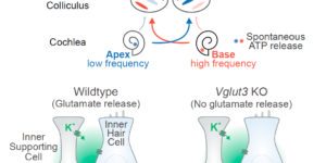
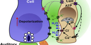
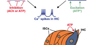
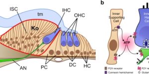
Purinergic Signaling in Cochlear Supporting Cells Reduces Hair Cell Excitability by Increasing the Extracellular Space

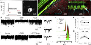
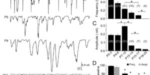
Neurons in developing sensory pathways exhibit spontaneous bursts of electrical activity that are critical for survival, maturation and circuit refinement. In the auditory system, intrinsically generated activity arises within the cochlea, but the molecular mechanisms that initiate this activity remain poorly understood. We show that burst firing of mouse inner hair cells prior to hearing onset requires P2RY1 autoreceptors expressed by inner supporting cells. P2RY1 activation triggers K+ efflux and depolarization of hair cells, as well as osmotic shrinkage of supporting cells that dramatically increased the extracellular space and speed of K+ redistribution. Pharmacological inhibition or genetic disruption of P2RY1 suppressed neuronal burst firing by reducing K+ release, but unexpectedly enhanced their tonic firing, as water resorption by supporting cells reduced the extracellular space, leading to K+ accumulation. These studies indicate that purinergic signaling in supporting cells regulates hair cell excitability by controlling the volume of the extracellular space.

Homeostatic Control of Spontaneous Activity in the Developing Auditory System.

Neurons in the developing auditory system exhibit spontaneous bursts of activity before hearing onset. How this intrinsically generated activity influences development remains uncertain, because few mechanistic studies have been performed in vivo. We show using macroscopic calcium imaging in unanesthetized mice that neurons responsible for processing similar frequencies of sound exhibit highly synchronized activity throughout the auditory system during this critical phase of development. Spontaneous activity normally requires synaptic excitation of spiral ganglion neurons (SGNs). Unexpectedly, tonotopic spontaneous activity was preserved in a mouse model of deafness in which glutamate release from hair cells is abolished. SGNs in these mice exhibited enhanced excitability, enabling direct neuronal excitation by supporting cell-induced potassium transients. These results indicate that homeostatic mechanisms maintain spontaneous activity in the pre-hearing period, with significant implications for both circuit development and therapeutic approaches aimed at treating congenital forms of deafness arising through mutations in key sensory transduction components.

NMDA Receptors Enhance Spontaneous Activity and Promote Neuronal Survival in the Developing Cochlea.

Spontaneous bursts of activity in developing sensory pathways promote maturation of neurons, refinement of neuronal connections, and assembly of appropriate functional networks. In the developing auditory system, inner hair cells (IHCs) spontaneously fire Ca(2+) spikes, each of which is transformed into a mini-burst of action potentials in spiral ganglion neurons (SGNs). Here we show that NMDARs are expressed in SGN dendritic terminals and play a critical role during transmission of activity from IHCs to SGNs before hearing onset. NMDAR activation enhances glutamate-mediated Ca(2+) influx at dendritic terminals, promotes repetitive firing of individual SGNs in response to each synaptic event, and enhances coincident activity of neighboring SGNs that will eventually encode similar frequencies of sound. Loss of NMDAR signaling from SGNs reduced their survival both in vivo and in vitro, revealing that spontaneous activity in the prehearing cochlea promotes maturation of auditory circuitry through periodic activation of NMDARs in SGNs.

Spontaneous Activity of Cochlear Hair Cells Triggered by Fluid Secretion Mechanism in Adjacent Support Cells.

Spontaneous electrical activity of neurons in developing sensory systems promotes their maturation and proper connectivity. In the auditory system, spontaneous activity of cochlear inner hair cells (IHCs) is initiated by the release of ATP from glia-like inner supporting cells (ISCs), facilitating maturation of central pathways before hearing onset. Here, we find that ATP stimulates purinergic autoreceptors in ISCs, triggering Cl(-) efflux and osmotic cell shrinkage by opening TMEM16A Ca(2+)-activated Cl(-) channels. Release of Cl(-) from ISCs also forces K(+) efflux, causing transient depolarization of IHCs near ATP release sites. Genetic deletion of TMEM16A markedly reduces the spontaneous activity of IHCs and spiral ganglion neurons in the developing cochlea and prevents ATP-dependent shrinkage of supporting cells. These results indicate that supporting cells in the developing cochlea have adapted a pathway used for fluid secretion in other organs to induce periodic excitation of hair cells.

Spontaneous activity in the developing auditory system.

Spontaneous electrical activity is a common feature of sensory systems during early development. This sensory-independent neuronal activity has been implicated in promoting their survival and maturation, as well as growth and refinement of their projections to yield circuits that can rapidly extract information about the external world. Periodic bursts of action potentials occur in auditory neurons of mammals before hearing onset. This activity is induced by inner hair cells (IHCs) within the developing cochlea, which establish functional connections with spiral ganglion neurons (SGNs) several weeks before they are capable of detecting external sounds. During this pre-hearing period, IHCs fire periodic bursts of Ca(2+) action potentials that excite SGNs, triggering brief but intense periods of activity that pass through auditory centers of the brain. Although spontaneous activity requires input from IHCs, there is ongoing debate about whether IHCs are intrinsically active and their firing periodically interrupted by external inhibitory input (IHC-inhibition model), or are intrinsically silent and their firing periodically promoted by an external excitatory stimulus (IHC-excitation model). There is accumulating evidence that inner supporting cells in Kölliker’s organ spontaneously release ATP during this time, which can induce bursts of Ca(2+) spikes in IHCs that recapitulate many features of auditory neuron activity observed in vivo. Nevertheless, the role of supporting cells in this process remains to be established in vivo. A greater understanding of the molecular mechanisms responsible for generating IHC activity in the developing cochlea will help reveal how these events contribute to the maturation of nascent auditory circuits.

Spontaneous regeneration of cochlear supporting cells after neonatal ablation ensures hearing in the adult mouse.

Supporting cells in the cochlea play critical roles in the development, maintenance, and function of sensory hair cells and auditory neurons. Although the loss of hair cells or auditory neurons results in sensorineural hearing loss, the consequence of supporting cell loss on auditory function is largely unknown. In this study, we specifically ablated inner border cells (IBCs) and inner phalangeal cells (IPhCs), the two types of supporting cells surrounding inner hair cells (IHCs) in mice in vivo. We demonstrate that the organ of Corti has the intrinsic capacity to replenish IBCs/IPhCs effectively during early postnatal development. Repopulation depends on the presence of hair cells and cells within the greater epithelial ridge and is independent of cell proliferation. This plastic response in the neonatal cochlea preserves neuronal survival, afferent innervation, and hearing sensitivity in adult mice. In contrast, the capacity for IBC/IPhC regeneration is lost in the mature organ of Corti, and consequently IHC survival and hearing sensitivity are impaired significantly, demonstrating that there is a critical period for the regeneration of cochlear supporting cells. Our findings indicate that the quiescent neonatal organ of Corti can replenish specific supporting cells completely after loss in vivo to guarantee mature hearing function.

Calcium action potentials in hair cells pattern auditory neuron activity before hearing onset.

We found rat central auditory neurons to fire action potentials in a precise sequence of mini-bursts before the age of hearing onset. This stereotyped pattern was initiated by hair cells in the cochlea, which trigger brief bursts of action potentials in auditory neurons each time they fire a Ca2+ spike. By generating theta-like activity, hair cells may limit the influence of synaptic depression in developing auditory circuits and promote consolidation of synapses.

ATP-induced morphological changes in supporting cells of the developing cochlea.

The developing cochlea of mammals contains a large group of columnar-shaped cells, which together form a structure known as Kölliker’s organ. Prior to the onset of hearing, these inner supporting cells periodically release adenosine 5′-triphosphate (ATP), which activates purinergic receptors in surrounding supporting cells, inner hair cells and the dendrites of primary auditory neurons. Recent studies indicate that purinergic signaling between inner supporting cells and inner hair cells initiates bursts of action potentials in auditory nerve fibers before the onset of hearing. ATP also induces prominent effects in inner supporting cells, including an increase in membrane conductance, a rise in intracellular Ca(2+), and dramatic changes in cell shape, although the importance of ATP signaling in non-sensory cells of the developing cochlea remains unknown. Here, we review current knowledge pertaining to purinergic signaling in supporting cells of Kölliker’s organ and focus on the mechanisms by which ATP induces changes in their morphology. We show that these changes in cell shape are preceded by increases in cytoplasmic Ca(2+), and provide new evidence indicating that elevation of intracellular Ca(2+) and IP(3) are sufficient to initiate shape changes. In addition, we discuss the possibility that these ATP-mediated morphological changes reflect crenation following the activation of Ca(2+)-activated Cl(-) channels, and speculate about the possible functions of these changes in cell morphology for maturation of the cochlea.

Developmental regulation of spontaneous activity in the Mammalian cochlea.

Neurons in the developing auditory system fire bursts of action potentials before the onset of hearing. This spontaneousactivity promotes the survival and maturation of auditory neurons and the refinement of synaptic connections in auditory nuclei; however, the mechanisms responsible for initiating this activity remain uncertain. Previous studies indicate that inner supporting cells (ISCs) in the developing cochlea periodically release ATP, which depolarizes inner hair cells (IHCs), leading to bursts of action potentials in postsynaptic spiral ganglion neurons (SGNs). To determine when purinergic signaling appears in the developing cochlea and whether it is responsible for initiating auditory neuron activity throughout the prehearing period, we examined spontaneousactivity from ISCs, IHCs, and SGNs in cochleae acutely isolated from rats during the first three postnatal weeks. We found that ATP was released from ISCs within the cochlea from birth until the onset of hearing, which led to periodic inward currents, Ca(2+) transients, and morphological changes in these supporting cells. This spontaneous release of ATP also depolarized IHCs and triggered bursts of action potentials in SGNs for most of the postnatal prehearing period, beginning a few days after birth as IHCs became responsive to ATP, until the onset of hearing when ATP was no longer released from ISCs. When IHCs were not subject to purinergic excitation, SGNs exhibited little or no activity. These results suggest that supporting cells in the cochlea provide the primary excitatory stimulus responsible for initiating bursts of action potentials in auditory nerve fibers before the onset of hearing.

The origin of spontaneous activity in the developing auditory system.

Spontaneous activity in the developing auditory system is required for neuronal survival as well as the refinement and maintenance of tonotopic maps in the brain. However, the mechanisms responsible for initiating auditory nerve firing in the absence of sound have not been determined. Here we show that supporting cells in the developing rat cochlea spontaneously release ATP, which causes nearby inner hair cells to depolarize and release glutamate, triggering discrete bursts of action potentials in primary auditory neurons. This endogenous, ATP-mediated signalling synchronizes the output of neighbouring inner hair cells, which may help refine tonotopic maps in the brain. Spontaneous ATP-dependent signalling rapidly subsides after the onset of hearing, thereby preventing this experience-independent activity from interfering with accurate encoding of sound. These data indicate that supporting cells in the organ of Corti initiate electrical activity in auditory nerves before hearing, pointing to an essential role for peripheral, non-sensory cells in the development of central auditory pathways.