Molecularly defined cortical astroglia subpopulation modulates neurons via secretion of Norrin.

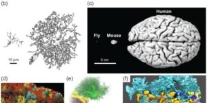
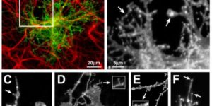
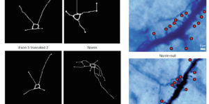
Despite expanding knowledge regarding the role of astroglia in regulating neuronal function, little is known about regional or functional subgroups of brain astroglia and how they may interact with neurons. We use an astroglia-specific promoter fragment in transgenic mice to identify an anatomically defined subset of adult gray matter astroglia. Using transcriptomic and histological analyses, we generate a combinatorial profile for the in vivo identification and characterization of this astroglia subpopulation. These astroglia are enriched in mouse cortical layer V; express distinct molecular markers, including Norrin and leucine-rich repeat-containing G-protein-coupled receptor 6 (LGR6), with corresponding layer-specific neuronal ligands; are found in the human cortex; and modulate neuronal activity. Astrocytic Norrin appears to regulate dendrites and spines; its loss, as occurring in Norrie disease, contributes to cortical dendritic spine loss. These studies provide evidence that human and rodent astroglia subtypes are regionally and functionally distinct, can regulate local neuronal dendrite and synaptic spine development, and contribute to disease.

Transient Opening of the Mitochondrial Permeability Transition Pore Induces Microdomain Calcium Transients in Astrocyte Processes.

Astrocytes extend highly branched processes that form functionally isolated microdomains, facilitating local homeostasis by redistributing ions, removing neurotransmitters, and releasing factors to influence blood flow and neuronal activity. Microdomains exhibit spontaneous increases in calcium (Ca2+), but the mechanisms and functional significance of this localized signaling are unknown. By developing conditional, membrane-anchored GCaMP3 mice, we found that microdomain activity that occurs in the absence of inositol triphosphate (IP3)-dependent release from endoplasmic reticulum arises through Ca2+ efflux from mitochondria during brief openings of the mitochondrial permeability transition pore. These microdomain Ca2+ transients were facilitated by the production of reactive oxygen species during oxidative phosphorylation and were enhanced by expression of a mutant form of superoxide dismutase 1 (SOD1 G93A) that causes astrocyte dysfunction and neurodegeneration in amyotrophic lateral sclerosis (ALS). By localizing mitochondria to microdomains, astrocytes ensure local metabolic support for energetically demanding processes and enable coupling between metabolic demand and Ca2+ signaling events.

Activity-dependent switch of GABAergic inhibition into glutamatergic excitation in astrocyte-neuron networks.

Interneurons are critical for proper neural network function and can activate Ca2+ signaling in astrocytes. However, the impact of the interneuron-astrocyte signaling into neuronal network operation remains unknown. Using the simplest hippocampal Astrocyte-Neuron network, i.e., GABAergic interneuron, pyramidal neuron, single CA3-CA1 glutamatergic synapse, and astrocytes, we found that interneuron-astrocyte signaling dynamically affected excitatory neurotransmission in an activity- and time-dependent manner, and determined the sign (inhibition vs potentiation) of the GABA-mediated effects. While synaptic inhibition was mediated by GABAA receptors, potentiation involved astrocyte GABAB receptors, astrocytic glutamate release, and presynaptic metabotropic glutamate receptors. Using conditional astrocyte-specific GABAB receptor (Gabbr1) knockout mice, we confirmed the glial source of the interneuron-induced potentiation, and demonstrated the involvement of astrocytes in hippocampal theta and gamma oscillations in vivo. Therefore, astrocytes decode interneuron activity and transform inhibitory into excitatory signals, contributing to the emergence of novel network properties resulting from the interneuron-astrocyte interplay.

Neuromodulators signal through astrocytes to alter neural circuit activity and behaviour.

Astrocytes associate with synapses throughout the brain and express receptors for neurotransmitters that can increase intracellular calcium (Ca2+). Astrocytic Ca2+ signalling has been proposed to modulate neural circuit activity, but the pathways that regulate these events are poorly defined and in vivo evidence linking changes in astrocyte Ca2+ levels to alterations in neurotransmission or behaviour is limited. Here we show that Drosophila astrocytes exhibit activity-regulated Ca2+ signalling in vivo. Tyramine and octopamine released from neurons expressing tyrosine decarboxylase 2 (Tdc2) signal directly to astrocytes to stimulate Ca2+ increases through the octopamine/tyramine receptor (Oct-TyrR) and the transient receptor potential (TRP) channel Water witch (Wtrw), and astrocytes in turn modulate downstream dopaminergic neurons. Application of tyramine or octopamine to live preparations silenced dopaminergic neurons and this inhibition required astrocytic Oct-TyrR and Wtrw. Increasing astrocyte Ca2+ signalling was sufficient to silence dopaminergic neuron activity, which was mediated by astrocyte endocytic function and adenosine receptors. Selective disruption of Oct-TyrR or Wtrw expression in astrocytes blocked astrocytic Ca2+ signalling and profoundly altered olfactory-driven chemotaxis and touch-induced startle responses. Our work identifies Oct-TyrR and Wtrw as key components of the astrocytic Ca2+ signalling machinery, provides direct evidence that octopamine- and tyramine-based neuromodulation can be mediated by astrocytes, and demonstrates that astrocytes are essential for multiple sensory-driven behaviours in Drosophila.

Large-scale recording of astrocyte activity.

Astrocytes are highly ramified glial cells found throughout the central nervous system (CNS). They express a variety of neurotransmitter receptors that can induce widespread chemical excitation, placing these cells in an optimal position to exert global effects on brain physiology. However, the activity patterns of only a small fraction of astrocytes have been examined and techniques to manipulate their behavior are limited. As a result, little is known about how astrocytes modulate CNS function on synaptic, microcircuit, or systems levels. Here, we review current and emerging approaches for visualizing and manipulating astrocyte activity in vivo. Deciphering how astrocyte network activity is controlled in different physiological and pathological contexts is crucial for defining their roles in the healthy and diseased CNS.

Human astrocytes develop physiological morphology and remain quiescent in a novel 3D matrix.

Astrocytes are the most abundant glial cells in the brain and are responsible for diverse functions, from modulating synapse function to regulating the blood-brain barrier. In vivo, these cells exhibit a star-shaped morphology with multiple radial processes that contact synapses and completely surround brain capillaries. In response to trauma or CNS disease, astrocytes become activated, a state associated with profound changes in gene expression, including upregulation of intermediate filament proteins, such as glial fibrillary acidic protein (GFAP). The inability to recapitulate the complex structure of astrocytes and maintain their quiescent state in vitro is a major roadblock to further developments in tissue engineering and regenerative medicine. Here, we characterize astrocyte morphology and activation in various hydrogels to assess the feasibility of developing a matrix that mimics key aspects of the native microenvironment. We show that astrocytes seeded in optimized matrix composed of collagen, hyaluronic acid, and matrigel exhibit a star-shaped morphology with radial processes and do not upregulate GFAP expression, hallmarks of quiescent astrocytes in the brain. In these optimized gels, collagen I provides structural support, HA mimics the brain extracellular matrix, and matrigel provides endothelial cell compatibility and was found to minimize GFAP upregulation. This defined 3D microenvironment for maintaining human astrocytes in vitro provides new opportunities for developing improved models of the blood-brain barrier and studying their response to stress signals.

Calcium dynamics in astrocyte processes during neurovascular coupling.

Enhanced neuronal activity in the brain triggers a local increase in blood flow, termed functional hyperemia, via several mechanisms, including calcium (Ca(2+)) signaling in astrocytes. However, recent in vivo studies have questioned the role of astrocytes in functional hyperemia because of the slow and sparse dynamics of their somatic Ca(2+) signals and the absence of glutamate metabotropic receptor 5 in adults. Here, we reexamined their role in neurovascular coupling by selectively expressing a genetically encoded Ca(2+) sensor in astrocytes of the olfactory bulb. We show that in anesthetized mice, the physiological activation of olfactory sensory neuron (OSN) terminals reliably triggers Ca(2+) increases in astrocyte processes but not in somata. These Ca(2+) increases systematically precede the onset of functional hyperemia by 1-2 s, reestablishing astrocytes as potential regulators of neurovascular coupling.

Norepinephrine controls astroglial responsiveness to local circuit activity.

Astrocytes perform crucial supportive functions, including neurotransmitter clearance, ion buffering, and metabolite delivery. They can also influence blood flow and neuronal activity by releasing gliotransmitters in response to intracellular Ca(2+) transients. However, little is known about how astrocytes are engaged during different behaviors in vivo. Here we demonstrate that norepinephrine primes astrocytes to detect changes in cortical network activity. We show in mice that locomotion triggers simultaneous activation of astrocyte networks in multiple brain regions. This global stimulation of astrocytes was inhibited by alpha-adrenoceptor antagonists and abolished by depletion of norepinephrine from the brain. Although astrocytes in visual cortex of awake mice were rarely engaged when neurons were activated by light stimulation alone, pairing norepinephrine release with light stimulation markedly enhanced astrocyte Ca(2+) signaling. Our findings indicate that norepinephrine shifts the gain of astrocyte networks according to behavioral state, enabling astrocytes to respond to local changes in neuronal activity.

Neuronal activity regulates glutamate transporter dynamics in developing astrocytes.

Glutamate transporters (GluTs) maintain a low ambient level of glutamate in the central nervous system (CNS) and shape the activation of glutamate receptors at synapses. Nevertheless, the mechanisms that regulate the trafficking and localization of transporters near sites of glutamate release are poorly understood. Here, we examined the subcellular distribution and dynamic remodeling of the predominant GluT GLT-1 (excitatory amino acid transporter 2, EAAT2) in developing hippocampal astrocytes. Immunolabeling revealed that endogenous GLT-1 is concentrated into discrete clusters along branches of developing astrocytes that were apposed preferentially to synapsin-1 positive synapses. Green fluorescent protein (GFP)-GLT-1 fusion proteins expressed in astrocytes also formed distinct clusters that lined the edges of astrocyte processes, as well as the tips of filopodia and spine-like structures. Time-lapse three-dimensional confocal imaging in tissue slices revealed that GFP-GLT-1 clusters were dynamically remodeled on a timescale of minutes. Some transporter clusters moved within developing astrocyte branches as filopodia extended and retracted, while others maintained stable positions at the tips of spine-like structures. Blockade of neuronalactivity with tetrodotoxin reduced both the density and perisynaptic localization of GLT-1 clusters. Conversely, enhancement of neuronal activity increased the size of GLT-1 clusters and their proximity to synapses. Together, these findings indicate that neuronal activity influences both the organization of GluTs in developing astrocyte membranes and their position relative to synapses.