Changes in the Excitability of Neocortical Neurons in a Mouse Model of Amyotrophic Lateral Sclerosis Are Not Specific to Corticospinal Neurons and Are Modulated by Advancing Disease

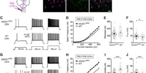
Cell type-specific changes in neuronal excitability have been proposed to contribute to the selective degeneration of corticospinal neurons in amyotrophic lateral sclerosis (ALS) and to neocortical hyperexcitability, a prominent feature of both inherited and sporadic variants of the disease, but the mechanisms underlying selective loss of specific cell types in ALS are not known. We analyzed the physiological properties of distinct classes of cortical neurons in the motor cortex of hSOD1G93A mice of both sexes and found that they all exhibit increases in intrinsic excitability that depend on disease stage. Targeted recordings and in vivo calcium imaging further revealed that neurons adapt their functional properties to normalize cortical excitability as the disease progresses. Although different neuron classes all exhibited increases in intrinsic excitability, transcriptional profiling indicated that the molecular mechanisms underlying these changes are cell type specific. The increases in excitability in both excitatory and inhibitory cortical neurons show that selective dysfunction of neuronal cell types cannot account for the specific vulnerability of corticospinal motor neurons in ALS. Furthermore, the stage-dependent alterations in neuronal function highlight the ability of cortical circuits to adapt as disease progresses. These findings show that both disease stage and cell type must be considered when developing therapeutic strategies for treating ALS.

Activity-dependent switch of GABAergic inhibition into glutamatergic excitation in astrocyte-neuron networks.

Interneurons are critical for proper neural network function and can activate Ca2+ signaling in astrocytes. However, the impact of the interneuron-astrocyte signaling into neuronal network operation remains unknown. Using the simplest hippocampal Astrocyte-Neuron network, i.e., GABAergic interneuron, pyramidal neuron, single CA3-CA1 glutamatergic synapse, and astrocytes, we found that interneuron-astrocyte signaling dynamically affected excitatory neurotransmission in an activity- and time-dependent manner, and determined the sign (inhibition vs potentiation) of the GABA-mediated effects. While synaptic inhibition was mediated by GABAA receptors, potentiation involved astrocyte GABAB receptors, astrocytic glutamate release, and presynaptic metabotropic glutamate receptors. Using conditional astrocyte-specific GABAB receptor (Gabbr1) knockout mice, we confirmed the glial source of the interneuron-induced potentiation, and demonstrated the involvement of astrocytes in hippocampal theta and gamma oscillations in vivo. Therefore, astrocytes decode interneuron activity and transform inhibitory into excitatory signals, contributing to the emergence of novel network properties resulting from the interneuron-astrocyte interplay.

Astrocyte glutamate transporters regulate metabotropic glutamate receptor-mediated excitation of hippocampal interneurons.

Clearance of extracellular glutamate is essential for limiting the activity of metabotropic glutamate receptors (mGluRs) at excitatory synapses; however, the relative contribution of transporters found in neuronal and glial membranes to this uptake is poorly understood. Hippocampalinterneurons located at the oriens-alveus border express mGluR1alpha, a metabotropic glutamate receptor that regulates excitability and synaptic plasticity. To determine which glutamate transporters are essential for removing glutamate at these excitatory synapses, we recorded mGluR1-mediated EPSCs from oriens-lacunosum moleculare (O-LM) interneurons in acute hippocampal slices. Stimulation in stratum oriens reliably elicited a slow mGluR1-mediated current in O-LM interneurons if they were briefly depolarized to allow Ca2+ entry before stimulation. Selective inhibition of GLT-1 [for glutamate transporter; EAAT2 (for excitatory amino acid transporter)] with dihydrokainate increased the amplitude of these responses approximately threefold, indicating that these transporters compete with mGluRs for synaptically released glutamate. However, inhibition of all glutamate transporters with TBOA (DL-threo-b-benzyloxyaspartic acid) increased mGluR1 EPSCs >15-fold, indicating that additional transporters also shape activation of these receptors. To identify these transporters, we examined mGluR1 EPSCs in mice lacking GLAST (for glutamate-aspartate transporter; EAAT1) or EAAC1 (for excitatory amino acid carrier; EAAT3). A comparison of responses recorded from wild-type and transporter knock-out mice revealed that the astroglial glutamate transporters GLT-1 and GLAST, but not the neuronal transporter EAAC1, restrict activation of mGluRs in O-LM interneurons. Transporter-dependent potentiation of mGluR1 EPSCs led to a dramatic increase in interneuron firing and enhanced inhibition of CA1 pyramidal neurons, suggesting that acute or prolonged disruption of transporter activity could lead to changes in network activity as a result of enhanced interneuron excitability.

Excitatory actions of norepinephrine on multiple classes of hippocampal CA1 interneurons

Norepinephrine (NE) causes an increase in the frequency of inhibitory postsynaptic potentials in CA1 pyramidal neurons in vitro. The possibility that this increase in tonic inhibition is caused by an excitatory effect on inhibitory interneurons was investigated through whole-cell recordings from pyramidal cells and both whole-cell and cell-attached patch recordings from visualized interneurons in acute slices of rat hippocampus. Adrenergic agonists caused a large increase in the frequency and amplitude of spontaneous IPSCs recorded from pyramidal cells in the presence of ionotropic glutamate receptor blockers, but they had no effect on either the frequency or the amplitude of action potential-independent miniature IPSCs recorded in tetrodotoxin. This effect was mediated primarily by an alpha adrenoceptor, although a slight beta adrenoceptor-dependent increase in IPSCs was also observed. NE caused interneurons located in all strata to depolarize and begin firing action potentials. Many of these cells had axons that ramified throughout the stratum pyramidale, suggesting that they are responsible for the IPSCs observed in pyramidal neurons. This depolarization was also mediated by an alpha adrenoceptor and was blocked by a selective alpha 1- but not a selective alpha 2-adrenoceptor antagonist. However, a slight beta adrenoceptor-dependent depolarization was detected in those interneurons that displayed time-dependent inward rectification. In the presence of a beta antagonist, NE induced an inward current that reversed near the predicted K+ equilibrium potential and was not affected by changes in intracellular Cl- concentration. In the presence of an alpha 1 antagonist, NE induced an inwardly rectifying current at potentials negative to approximately -70 mV that did not reverse (between -130 and -60 mV), characteristics similar to the hyperpolarization-activated current (lh). However, the depolarizing action of NE is attributable primarily to the alpha 1 adrenoceptor-mediated decrease in K+ conductance and not the beta adrenoceptor-dependent increase in lh. These results provide evidence that NE increases action potential-dependent IPSCs in pyramidal neurons by depolarizing surrounding inhibitory interneurons. This potent excitatory action of NE on multiple classes of hippocampal interneurons may contribute to the NE-induced decrease in the spontaneous activity of pyramidal neurons and the antiepileptic effects of NE observed in vivo.