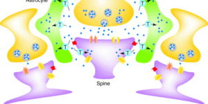
The density of EAAC1 (EAAT3) glutamate transporters expressed by neurons in the mammalian CNS.

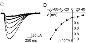
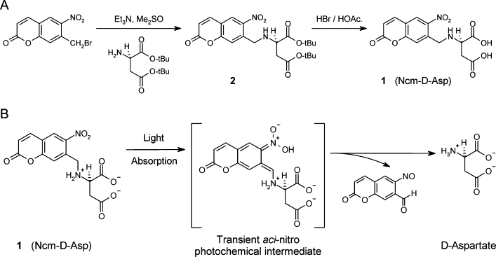
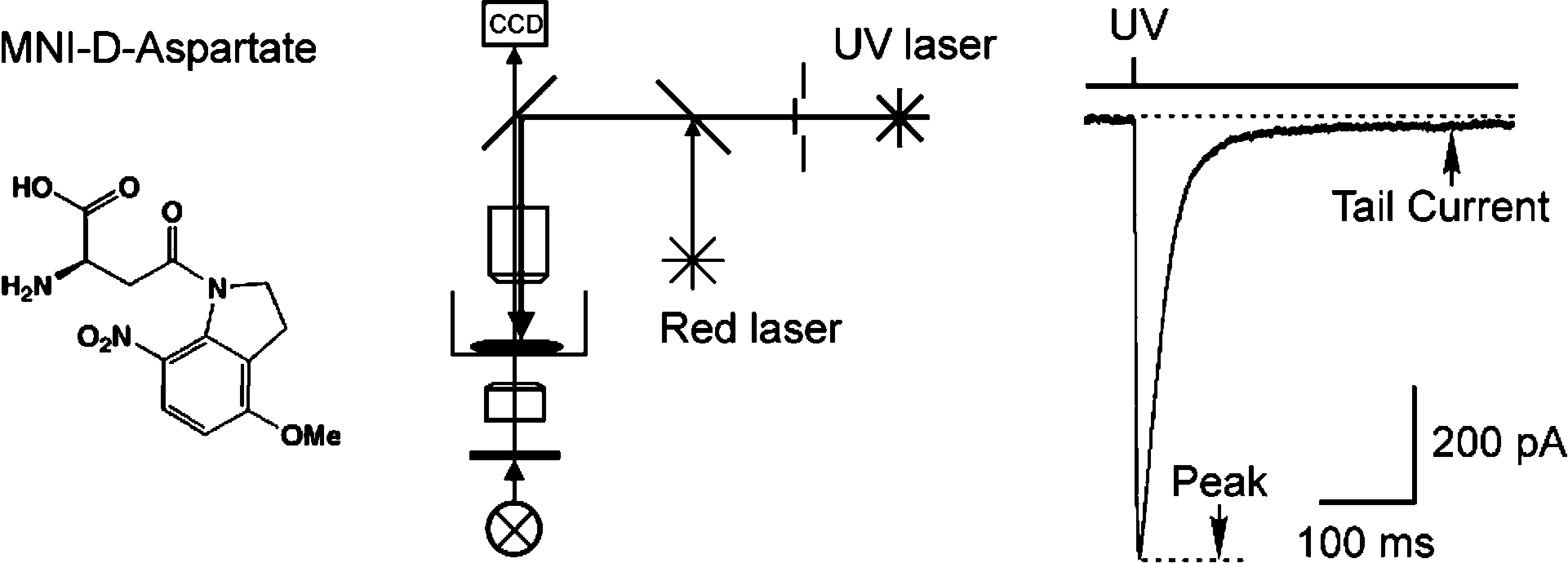
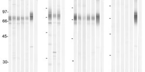
The extracellular levels of excitatory amino acids are kept low by the action of the glutamate transporters. Glutamate/aspartate transporter (GLAST) and glutamate transporter-1 (GLT-1) are the most abundant subtypes and are essential for the functioning of the mammalian CNS, but the contribution of the EAAC1 subtype in the clearance of synaptic glutamate has remained controversial, because the density of this transporter in different tissues has not been determined. We used purified EAAC1 protein as a standard during immunoblotting to measure the concentration of EAAC1 in different CNS regions. The highest EAAC1 levels were found in the young adult rat hippocampus. Here, the concentration of EAAC1 was ∼0.013 mg/g tissue (∼130 molecules μm⁻³), 100 times lower than that of GLT-1. Unlike GLT-1 expression, which increases in parallel with circuit formation, only minor changes in the concentration of EAAC1 were observed from E18 to adulthood. In hippocampal slices, photolysis of MNI-D-aspartate (4-methoxy-7-nitroindolinyl-D-aspartate) failed to elicit EAAC1-mediated transporter currents in CA1 pyramidal neurons, and D-aspartate uptake was not detected electron microscopically in spines. Using EAAC1 knock-out mice as negative controls to establish antibody specificity, we show that these relatively small amounts of EAAC1 protein are widely distributed in somata and dendrites of all hippocampal neurons. These findings raise new questions about how so few transporters can influence the activation of NMDA receptors at excitatory synapses.

Neuronal activity regulates glutamate transporter dynamics in developing astrocytes.

Glutamate transporters (GluTs) maintain a low ambient level of glutamate in the central nervous system (CNS) and shape the activation of glutamate receptors at synapses. Nevertheless, the mechanisms that regulate the trafficking and localization of transporters near sites of glutamate release are poorly understood. Here, we examined the subcellular distribution and dynamic remodeling of the predominant GluT GLT-1 (excitatory amino acid transporter 2, EAAT2) in developing hippocampal astrocytes. Immunolabeling revealed that endogenous GLT-1 is concentrated into discrete clusters along branches of developing astrocytes that were apposed preferentially to synapsin-1 positive synapses. Green fluorescent protein (GFP)-GLT-1 fusion proteins expressed in astrocytes also formed distinct clusters that lined the edges of astrocyte processes, as well as the tips of filopodia and spine-like structures. Time-lapse three-dimensional confocal imaging in tissue slices revealed that GFP-GLT-1 clusters were dynamically remodeled on a timescale of minutes. Some transporter clusters moved within developing astrocyte branches as filopodia extended and retracted, while others maintained stable positions at the tips of spine-like structures. Blockade of neuronalactivity with tetrodotoxin reduced both the density and perisynaptic localization of GLT-1 clusters. Conversely, enhancement of neuronal activity increased the size of GLT-1 clusters and their proximity to synapses. Together, these findings indicate that neuronal activity influences both the organization of GluTs in developing astrocyte membranes and their position relative to synapses.

Zones of enhanced glutamate release from climbing fibers in the mammalian cerebellum.

Purkinje cells in the mammalian cerebellum are remarkably homogeneous in shape and orientation, yet they exhibit regional differences in gene expression. Purkinje cells that express high levels of zebrin II (aldolase C) and the glutamate transporter EAAT4 cluster in parasagittal zones that receive input from distinct groups of climbing fibers (CFs); however, the physiological properties of CFs that target these molecularly distinct Purkinje cells have not been determined. Here we report that CFs that innervate Purkinje cells in zebrin II-immunoreactive (Z(+)) zones release more glutamate per action potential than CFs in Z(-) zones. CF terminals in Z(+) zones had larger pools of release-ready vesicles, exhibited enhanced multivesicular release, and produced larger synaptic glutamate transients. As a result, CF-mediated EPSCs in Purkinje cells decayed more slowly in Z(+) zones, which triggered longer-duration complex spikes containing a greater number of spikelets. The differences in the duration of CF EPSCs between Z(+) and Z(-) zones persisted in EAAT4 knock-out mice, indicating that EAAT4 is not required for maintaining this aspect of CF function. These results indicate that the organization of the cerebellum into discrete longitudinal zones is defined not only by molecular phenotype of Purkinje cells within zones, but also by the physiological properties of CFs that project to these distinct regions. The enhanced release of glutamate from CFs in Z(+) zones may alter the threshold for synaptic plasticity and prolong inhibition of cerebellar output neurons in deep cerebellar nuclei.

Variations in promoter activity reveal a differential expression and physiology of glutamate transporters by glia in the developing and mature CNS

Glutamate transporters regulate excitatory neurotransmission and prevent glutamate-mediated excitotoxicity in the CNS. To better study the cellular and temporal dynamics of the expression of these transporters, we generated bacterial artificial chromosome promoter Discosoma red [glutamate-aspartate transporter (GLAST)] and green fluorescent protein [glutamate transporter-1 (GLT-1)] reporter transgenic mice. Analysis of these mice revealed a differential activation of the transporter promoters not previously appreciated. GLT-1 promoter activity in the adult CNS is almost completely restricted to astrocytes, often and unexpectedly in a nonoverlapping pattern with GLAST. Spinal cord GLT-1 promoter reporter, protein density, and physiology were 10-fold lower than in brain, suggesting a possible mechanism for regional sensitivity seen in disease. The GLAST promoter is active in both radial glia and many astrocytes in the developing CNS but is downregulated in most astrocytes as the mice mature. In the adult CNS, the highest GLAST promoter activity was observed in radial glia, such as those located in the subgranular layer of the dentate gyrus. The continued expression of GLAST by these neural progenitors raises the possibility that GLAST may have an unanticipated role in regulating their behavior. In addition, GLAST promoter activation was observed in oligodendrocytes in white matter throughout many (e.g., spinal cord and corpus callosum), but not all (e.g., cerebellum), CNS fiber tracts. Overall, these studies of GLT-1 and GLAST promoter activity, protein expression, and glutamate uptake revealed a close correlation between transgenic reporter signals and uptake capacity, indicating that these mice provide the means to monitor the expression and regulation of glutamate transporters in situ.

Analysis of cerebellar Purkinje cells using EAAT4 glutamate transporter promoter reporter in mice generated via bacterial artificial chromosome-mediated transgenesis

The EAAT4 glutamate transporter helps regulate excitatory neurotransmission and prevents glutamate-mediated excitotoxicity in the cerebellum. Immunohistochemistry and in situ hybridization have previously defined a cerebellar cell population expressing this protein. These methods, however, are not well suited for evaluating the dynamic regulation of the transporter and its gene-especially in living tissues. To better study EAAT4 expression and regulation, we generated bacterial artificial chromosome (BAC) promoter eGFP reporter transgenic mice. Histological analysis of the transgenic mice revealed that the EAAT4 promoter is active predominantly in Purkinje cells, but can also be modestly detected in other neurons early postnatally. EAAT4 promoter activity was not present in non-neuronal cells. Cerebellar organotypic slice cultures prepared from BAC transgenic mice provided a unique reagent to study transporter and Purkinje cell expression and regulation in living tissue. The correlation of promoter activity to protein expression makes the EAAT4 BAC promoter reporter a valuable tool to study regulation of EAAT4 expression.

The glutamate-aspartate transporter GLAST mediates glutamate uptake at inner hair cell afferent synapses in the mammalian cochlea.

Ribbon synapses formed between inner hair cells (IHCs) and afferent dendrites in the mammalian cochlea can sustain high rates of release, placing strong demands on glutamate clearance mechanisms. To investigate the role of transporters in glutamate removal at these synapses, we made whole-cell recordings from IHCs, afferent dendrites, and glial cells adjacent to IHCs [inner phalangeal cells (IPCs)] in whole-mount preparations of rat organ of Corti. Focal application of the transporter substrate D-aspartate elicited inward currents in IPCs, which were larger in the presence of anions that permeate the transporter-associated anion channel and blocked by the transporter antagonist D,L-threo-beta-benzyloxyaspartate. These currents were produced by glutamate-aspartate transporters (GLAST) (excitatory amino acid transporter 1) because they were weakly inhibited by dihydrokainate, an antagonist of glutamate transporter-1 (excitatory amino acid transporter 2) and were absent from IPCs in GLAST-/- cochleas. Furthermore, D-aspartate-induced currents in outside-out patches from IPCs exhibited larger steady-state currents than responses elicited by L-glutamate, a prominent feature of GLAST, and examination of cochlea from GLAST-Discosoma red (DsRed) promoter reporter mice revealed that DsRed expression was restricted to IPCs and other supporting cells surrounding IHCs. Saturation of transporters by photolysis of caged D-aspartate failed to elicit transporter currents in IHCs, as did local application of D-aspartate to afferent terminals, indicating that neither presynaptic nor postsynaptic membranes are major sites for glutamate removal. These data indicate that GLAST in supporting cells is responsible for transmitter uptake at IHC afferent synapses.

Ncm-D-aspartate: a novel caged D-aspartate suitable for activation of glutamate transporters and N-methyl-D-aspartate (NMDA) receptors in brain tissue

The D-isomer of aspartate is both a substrate for glutamate transporters and an agonist of N-methyl-D-aspartate (NMDA) receptors. To monitor the behavior of these receptors and transporters in intact tissue we synthesized a new photo-labile analogue of D-aspartate, N-[(6-nitrocoumarin-7-yl)methyl]-D-aspartic acid (Ncm-D-aspartate). This compound was photolyzed rapidly (t(1/2)=0.11 micros) by UV light with a quantum efficiency of 0.041 at pH 7.4. In acute hippocampal slices, photolysis of Ncm-D-aspartate by brief (1 ms) exposure to UV light elicited rapidly activating inward currents in astrocytes that were sensitive to inhibition by the glutamate transporter antagonist DL-threo-beta-benzyloxyaspartic acid (TBOA). Neither Ncm-D-aspartate nor the photo-released caging group exhibited agonist or antagonist activity at glutamate transporters, and Ncm-D-aspartate did not induce transporter currents prior to photolysis. Glutamate transporter currents were also elicited in cerebellar Purkinje cells in response to photolysis of Ncm-D-aspartate. Photo-release of D-aspartate from Ncm-D-aspartate did not induce alpha-amino-3-hydroxy-5-methyl-4-isoxazole propionic acid (AMPA)/kainate receptor or metabotropic glutamate receptor (mGluR) currents, but triggered robust NMDA receptor currents in neurons; Ncm-D-aspartate and the photolzyed caging group were similarly inert at NMDA receptors. These results indicate that Ncm-D-aspartate can be used to study NMDA receptors at excitatory synapses and interactions between transporters and receptors in brain tissue.

Synthesis and characterization of 4-methoxy-7-nitroindolinyl-D-aspartate, a caged compound for selective activation of glutamate transporters and N-methyl-D-aspartate receptors in brain tissue.

The D-isomer of aspartate is efficiently transported by high-affinity Na(+)/K(+)-dependent glutamate transporters and is an effective ligand of N-methyl-d-aspartate (NMDA) receptors. To facilitate analysis of the regulation of these proteins in their native membranes, we synthesized a photolabile analogue of D-aspartate, 4-methoxy-7-nitroindolinyl-D-aspartate (MNI-D-aspartate). This compound was photolyzed with a quantum efficiency of 0.09 at pH 7.4. Photorelease of d-aspartate in acute hippocampal slices through brief (1 ms) UV laser illumination of MNI-d-aspartate triggered rapidly activating currents in astrocytes that were inhibited by the glutamate transporter antagonist DL-threo-beta-benzyloxyaspartic acid (TBOA), indicating that they resulted from electrogenic uptake of D-aspartate. These transporter currents exhibited a distinct tail component that was approximately 2% of the peak current, which may result from the release of K(+) into the extracellular space during counter transport. MNI-D-aspartate was neither an agonist nor an antagonist of glutamate transporters at concentrations up to 500 muM and was stable in aqueous solution for several days. Glutamate transporter currents were also elicited in Bergmann glial cells and Purkinje neurons of the cerebellum in response to photolysis of MNI-D-aspartate, indicating that this compound can be used for monitoring the occupancy and regulation of glutamate transporters in different brain regions. Photorelease of D-aspartate did not activate alpha-amino-3-hydroxy-5-methyl-4-isoxazolepropionic acid (AMPA)/kainate receptors or metabotropic glutamate receptors (mGluRs) in neurons, but resulted in the selective, but transient, activation of NMDA receptors in hippocampal pyramidal neurons; MNI-D-aspartate was not an antagonist of NMDA receptors. These results indicate that MNI-D-aspartate also may be useful for studying the regulation of NMDA receptors at excitatory synapses.

Beta-lactam antibiotics offer neuroprotection by increasing glutamate transporter expression.

Glutamate is the principal excitatory neurotransmitter in the nervous system. Inactivation of synaptic glutamate is handled by the glutamate transporter GLT1 (also known as EAAT2; refs 1, 2), the physiologically dominant astroglial protein. In spite of its critical importance in normal and abnormal synaptic activity, no practical pharmaceutical can positively modulate this protein. Animal studies show that the protein is important for normal excitatory synaptic transmission, while its dysfunction is implicated in acute and chronic neurological disorders, including amyotrophic lateral sclerosis (ALS), stroke, brain tumours and epilepsy. Using a blinded screen of 1,040 FDA-approved drugs and nutritionals, we discovered that many beta-lactam antibiotics are potent stimulators of GLT1 expression. Furthermore, this action appears to be mediated through increased transcription of the GLT1 gene. beta-Lactams and various semi-synthetic derivatives are potent antibiotics that act to inhibit bacterial synthetic pathways. When delivered to animals, the beta-lactam ceftriaxone increased both brain expression of GLT1 and its biochemical and functional activity. Glutamate transporters are important in preventing glutamate neurotoxicity. Ceftriaxone was neuroprotective in vitro when used in models of ischaemic injury and motor neuron degeneration, both based in part on glutamate toxicity. When used in an animal model of the fatal disease ALS, the drug delayed loss of neurons and muscle strength, and increased mouse survival. Thus these studies provide a class of potential neurotherapeutics that act to modulate the expression of glutamate neurotransmitter transporters via gene activation.

Glutamate transporters bring competition to the synapse

Glutamate transporters (GluTs) prevent the accumulation of glutamate and influence the occupancy of receptors at synapses. The ability of extrasynaptic NMDA receptors and metabotropic glutamate receptors to participate in signaling is tightly regulated by GluT activity. Astrocytes express the highest density of GluTs and dominate clearance away from these receptors; synapses that are not associated with astrocyte processes experience greater mGluR activation and can be exposed to glutamate released at adjacent synapses. Although less abundant, neuronal transporters residing in the postsynaptic membrane can also shield receptors from the glutamate that is released. The diversity in synaptic morphology suggests a correspondingly rich diversity of GluT function in excitatory transmission.