Astrocyte glutamate transporters regulate metabotropic glutamate receptor-mediated excitation of hippocampal interneurons.

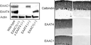
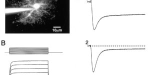
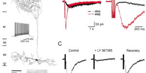
Clearance of extracellular glutamate is essential for limiting the activity of metabotropic glutamate receptors (mGluRs) at excitatory synapses; however, the relative contribution of transporters found in neuronal and glial membranes to this uptake is poorly understood. Hippocampalinterneurons located at the oriens-alveus border express mGluR1alpha, a metabotropic glutamate receptor that regulates excitability and synaptic plasticity. To determine which glutamate transporters are essential for removing glutamate at these excitatory synapses, we recorded mGluR1-mediated EPSCs from oriens-lacunosum moleculare (O-LM) interneurons in acute hippocampal slices. Stimulation in stratum oriens reliably elicited a slow mGluR1-mediated current in O-LM interneurons if they were briefly depolarized to allow Ca2+ entry before stimulation. Selective inhibition of GLT-1 [for glutamate transporter; EAAT2 (for excitatory amino acid transporter)] with dihydrokainate increased the amplitude of these responses approximately threefold, indicating that these transporters compete with mGluRs for synaptically released glutamate. However, inhibition of all glutamate transporters with TBOA (DL-threo-b-benzyloxyaspartic acid) increased mGluR1 EPSCs >15-fold, indicating that additional transporters also shape activation of these receptors. To identify these transporters, we examined mGluR1 EPSCs in mice lacking GLAST (for glutamate-aspartate transporter; EAAT1) or EAAC1 (for excitatory amino acid carrier; EAAT3). A comparison of responses recorded from wild-type and transporter knock-out mice revealed that the astroglial glutamate transporters GLT-1 and GLAST, but not the neuronal transporter EAAC1, restrict activation of mGluRs in O-LM interneurons. Transporter-dependent potentiation of mGluR1 EPSCs led to a dramatic increase in interneuron firing and enhanced inhibition of CA1 pyramidal neurons, suggesting that acute or prolonged disruption of transporter activity could lead to changes in network activity as a result of enhanced interneuron excitability.

Climbing fiber activation of EAAT4 transporters and kainate receptors in cerebellar Purkinje cells

Cerebellar Purkinje cells (PCs) express two glutamate transporters, EAAC1 (EAAT3) and EAAT4; however, their relative contribution to the uptake of glutamate at synapses is not known. We found that glutamate transporter currents recorded at climbing fiber (CF)-PC synapses are absent in mice lacking EAAT4 but unchanged in mice lacking EAAC1, indicating that EAAT4 is preferentially involved in clearing glutamate from CF synapses. However, comparison of CF synaptic currents between wild-type and transporter knock-out mice revealed that ionotropic glutamate receptors are responsible for >40% of the current previously attributed to transporters, indicating that PCs remove <10% of the glutamate released by the CF. The receptors responsible for the nontransporter component accounted for 5% of the CF EPSC, had a slower time course and lower occupancy than AMPA receptors at CF synapses, and exhibited pharmacological properties consistent with kainate receptors. In GluR5 knock-out mice, this current was dramatically reduced, indicating that CF excitation of PCs involves two distinct classes of ionotropic glutamate receptors, AMPA receptors and GluR5-containing kainate receptors.

Glial contribution to glutamate uptake at Schaffer collateral-commissural synapses in the hippocampus

Astrocytes in the hippocampus express high-affinity glutamate transporters that are important for lowering the concentration of extracellular glutamate after release at excitatory synapses. These transporters exhibit a permeability to chaotropic anions that is associated with transport, allowing their activity to be monitored in cell-fee patches when highly permeant anions are present. Astrocyte glutamate transporters are highly temperature sensitive, because L-glutamate-activated, anion-potentiated transporter currents in outside-out patches from these cells exhibited larger amplitudes and faster kinetics at 36 degreesC than at 24 degreesC. The cycling rate of these transporters was estimated by using paired applications of either L-glutamate or D-aspartate to measure the time necessary for the peak of the transporter current to recover from the steady-state level. Transporter currents in patches recovered with a time constant of 11.6 msec at 36 degreesC, suggesting that either the turnover rate of native transporters is much faster than previously reported for expressed EAAT2 transporters or the efficiency of these transporters is very low. Synaptically activated transporter currents persisted in astrocytes at physiological temperatures, although no evidence of these currents was found in CA1 pyramidal neurons in response to afferent stimulation. L-glutamate-gated transporter currents were also not detected in outside-out patches from pyramidal neurons. These results are consistent with the hypothesis that astrocyte transporters are responsible for taking up the majority of glutamate released at Schaffer collateral-commissural synapses in the hippocampus.Glial contribution to glutamate uptake at Schaffer collateral-commissural synapses in the hippocampus

Glutamate Release Monitored with Astrocyte Transporter Currents during LTP

Long-term potentiation (LTP) of synaptic transmission in the CA1 region of the hippocampus is thought to result from either increased transmitter release, heightened postsynaptic sensitivity, or a combination of the two. We have measured evoked glutamate release from Schaffer collateral/commissural fiber terminals in CA1 by recording synaptically activated glutamate transporter currents in hippocampal astrocytes located in stratum radiatum. Although several manipulations of release probability caused parallel changes in extracellular field potentials and synaptically activated transporter current amplitudes, induction of LTP failed to alter transporter-mediated responses, suggesting that LTP does not alter the amount of glutamate released upon synaptic stimulation.

Glutamate transporter currents in Bergmann glial cells follow the time course of extrasynaptic glutamate.

Glutamate transporters in the central nervous system are expressed in both neurons and glia, they mediate high affinity, electrogenic uptake of glutamate, and they are associated with an anion conductance that is stoichiometrically uncoupled from glutamate flux. Although a complete cycle of transport may require 50-100 ms, previous studies suggest that transporters can alter synaptic currents on a much faster time scale. We find that application of L-glutamate to outside-out patches from cerebellar Bergmann glia activates anion-potentiated glutamate transporter currents that activate in <1 ms, suggesting an efficient mechanism for the capture of extrasynaptic glutamate. Stimulation in the granule cell layer in cerebellar slices elicits all or none alpha-amino-3-hydroxy-5-methyl-4-isoxazolepropionate receptor and glutamate transporter currents in Bergmann glia that have a rapid onset, suggesting that glutamate released from climbing fiber terminals escapes synaptic clefts and reaches glial membranes shortly after release. Comparison of the concentration dependence of both alpha-amino-3-hydroxy-5-methyl-4-isoxazolepropionate receptor and glutamate transporter kinetics in patches with the time course of climbing fiber-evoked responses indicates that the glutamate transient at Bergmann glial membranes reaches a lower concentration than attained in the synaptic cleft and remains elevated in the extrasynaptic space for many milliseconds.

Synaptic activation of glutamate transporters in hippocampal astrocytes

Glutamate transporters in the CNS are expressed in neurons and glia and mediate high affinity, electrogenic uptake of extracellular glutamate. Although glia have the highest capacity for glutamate uptake, the amount of glutamate that reaches glial membranes following release and the rate that glial transporters bind and sequester transmitter is not known. We find that stimulation of Schaffer collateral/commissural fibers in hippocampal slices evokes glutamate transporter currents in CA1 astrocytes that activate rapidly, indicating that a significant amount of transmitter escapes the synaptic cleft shortly after release. Transporter currents in outside-out patches from astrocytes have faster kinetics than synaptically elicited currents, suggesting that the glutamate concentration attained at astrocytic membranes is lower but remains elevated for longer than in the synaptic cleft.