Oligodendrocytes control potassium accumulation in white matter and seizure susceptibility

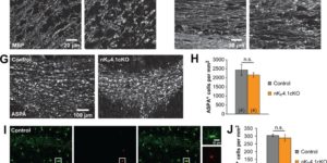
The inwardly rectifying K+ channel Kir4.1 is broadly expressed by CNS glia and deficits in Kir4.1 lead to seizures and myelin vacuolization. However, the role of oligodendrocyte Kir4.1 channels in controlling myelination and K+ clearance in white matter has not been defined. Here, we show that selective deletion of Kir4.1 from oligodendrocyte progenitors (OPCs) or mature oligodendrocytes did not impair their development or disrupt the structure of myelin. However, mice lacking oligodendrocyte Kir4.1 channels exhibited profound functional impairments, including slower clearance of extracellular K+ and delayed recovery of axons from repetitive stimulation in white matter, as well as spontaneous seizures, a lower seizure threshold, and activity-dependent motor deficits. These results indicate that Kir4.1 channels in oligodendrocytes play an important role in extracellular K+ homeostasis in white matter, and that selective loss of this channel from oligodendrocytes is sufficient to impair K+ clearance and promote seizures.
Electrophysiological properties of NG2(+) cells: Matching physiological studies with gene expression profiles.

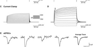
NG2(+) glial cells are a dynamic population of non-neuronal cells that give rise to myelinating oligodendrocytes in the central nervous system. These cells express numerous ion channels and neurotransmitter receptors, which endow them with a complex electrophysiological profile that is unique among glial cells. Despite extensive analysis of the electrophysiological properties of these cells, relatively little was known about the molecular identity of the channels and receptors that they express. The generation of new RNA-Seq datasets for NG2(+) cells has provided the means to explore how distinct genes contribute to the physiological properties of these progenitors. In this review, we systematically compare the results obtained through RNA-Seq transcriptional analysis of purified NG2(+) cells to previous physiological and molecular studies of these cells to define the complement of ion channels and neurotransmitter receptors expressed by NG2(+) cells in the mammalian brain and discuss the potential significance of the unique physiological properties of these cells.

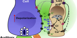
Spontaneous Activity of Cochlear Hair Cells Triggered by Fluid Secretion Mechanism in Adjacent Support Cells.

Spontaneous electrical activity of neurons in developing sensory systems promotes their maturation and proper connectivity. In the auditory system, spontaneous activity of cochlear inner hair cells (IHCs) is initiated by the release of ATP from glia-like inner supporting cells (ISCs), facilitating maturation of central pathways before hearing onset. Here, we find that ATP stimulates purinergic autoreceptors in ISCs, triggering Cl(-) efflux and osmotic cell shrinkage by opening TMEM16A Ca(2+)-activated Cl(-) channels. Release of Cl(-) from ISCs also forces K(+) efflux, causing transient depolarization of IHCs near ATP release sites. Genetic deletion of TMEM16A markedly reduces the spontaneous activity of IHCs and spiral ganglion neurons in the developing cochlea and prevents ATP-dependent shrinkage of supporting cells. These results indicate that supporting cells in the developing cochlea have adapted a pathway used for fluid secretion in other organs to induce periodic excitation of hair cells.