Changes in the Oligodendrocyte Progenitor Cell Proteome With Ageing

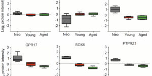
Following central nervous system (CNS) demyelination, adult oligodendrocyte progenitor cells (OPCs) can differentiate into new myelin-forming oligodendrocytes in a regenerative process called remyelination. While remyelination is very efficient in young adults, its efficiency declines progressively with ageing. Here we performed proteomic analysis of OPCs freshly isolated from the brains of neonate, young and aged female rats. Approximately 50% of the proteins are expressed at different levels in OPCs from neonates compared to their adult counterparts. The amount of myelin-associated proteins, and proteins associated with oxidative phosphorylation, inflammatory responses and actin cytoskeletal organization increased with age, while cholesterol-biosynthesis, transcription factors and cell cycle proteins decreased. Our experiments provide the first ageing OPC proteome, revealing the distinct features of OPCs at different ages. These studies provide new insights into why remyelination efficiency declines with ageing and potential roles for aged OPCs in other neurodegenerative diseases.

Oligodendrocyte precursor cells present antigen and are cytotoxic targets in inflammatory demyelination.

Oligodendrocyte precursor cells (OPCs) are abundant in the adult central nervous system, and have the capacity to regenerate oligodendrocytes and myelin. However, in inflammatory diseases such as multiple sclerosis (MS) remyelination is often incomplete. To investigate how neuroinflammation influences OPCs, we perform in vivo fate-tracing in an inflammatory demyelinating mouse model. Here we report that OPC differentiation is inhibited by both effector T cells and IFNγ overexpression by astrocytes. IFNγ also reduces the absolute number of OPCs and alters remaining OPCs by inducing the immunoproteasome and MHC class I. In vitro, OPCs exposed to IFNγ cross-present antigen to cytotoxic CD8 T cells, resulting in OPC death. In human demyelinated MS brain lesions, but not normal appearing white matter, oligodendroglia exhibit enhanced expression of the immunoproteasome subunit PSMB8. Therefore, OPCs may be co-opted by the immune system in MS to perpetuate the autoimmune response, suggesting that inhibiting immune activation of OPCs may facilitate remyelination.

Myelinogenic Plasticity of Oligodendrocyte Precursor Cells following Spinal Cord Contusion Injury

Spontaneous remyelination occurs after spinal cord injury (SCI), but the extent of myelin repair and identity of the cells responsible remain incompletely understood and contentious. We assessed the cellular origin of new myelin by fate mapping platelet-derived growth factor receptor α (PDGFRα), Olig2+, and P0+ cells following contusion SCI in mice. Oligodendrocyte precursor cells (OPCs; PDGFRα+) produced oligodendrocytes responsible for de novo ensheathment of ∼30% of myelinated spinal axons at injury epicenter 3 months after SCI, demonstrating that these resident cells are a major contributor to oligodendrocyte regeneration. OPCs also produced the majority of myelinating Schwann cells in the injured spinal cord; invasion of peripheral myelinating (P0+) Schwann cells made only a limited contribution. These findings reveal that PDGFRα+ cells perform diverse roles in CNS repair, as multipotential progenitors that generate both classes of myelinating cells. This endogenous repair might be exploited as a therapeutic target for CNS trauma and disease.

Cell-type specific differences in promoter activity of the ALS-linked C9orf72 mouse ortholog

A hexanucleotide repeat expansion in the C9orf72 gene is the most common cause of inherited forms of the neurodegenerative disease amyotrophic lateral sclerosis (ALS). Both loss-of-function and gain-of-function mechanisms have been proposed to underlie this disease, but the pathogenic pathways are not fully understood. To better understand the involvement of different cell types in the pathogenesis of ALS, we systematically analyzed the distribution of promoter activity of the mouse ortholog of C9orf72 in the central nervous system. We demonstrate that C9orf72 promoter activity is widespread in both excitatory and inhibitory neurons as well as in oligodendrocytes and oligodendrocyte precursor cells. In contrast, few microglia and astrocytes exhibit detectable C9orf72 promoter activity. Although at a gross level, the distribution of C9orf72 promoter activity largely follows overall cellular density, we found that it is selectively enriched in subsets of neurons and glial cells that degenerate in ALS. Specifically, we show that C9orf72 promoter activity is enriched in corticospinal and spinal motor neurons as well as in oligodendrocytes in brain regions that are affected in ALS. These results suggest that cell autonomous changes in both neurons and glia may contribute to C9orf72-mediated disease, as has been shown for mutations in superoxide dismutase-1 (SOD1).

Oligodendrocyte Development and Plasticity.

Oligodendrocyte precursor cells (OPCs) originate in the ventricular zones (VZs) of the brain and spinal cord and migrate throughout the developing central nervous system (CNS) before differentiating into myelinating oligodendrocytes (OLs). It is not known whether OPCs or OLs from different parts of the VZ are functionally distinct. OPCs persist in the postnatal CNS, where they continue to divide and generate myelinating OLs at a decreasing rate throughout adult life in rodents. Adult OPCs respond to injury or disease by accelerating their cell cycle and increasing production of OLs to replace lost myelin. They also form synapses with unmyelinated axons and respond to electrical activity in those axons by generating more OLs and myelin locally. This experience-dependent “adaptive” myelination is important in some forms of plasticity and learning, for example, motor learning. We review the control of OL lineage development, including OL population dynamics and adaptive myelination in the adult CNS.

Electrophysiological properties of NG2(+) cells: Matching physiological studies with gene expression profiles.

NG2(+) glial cells are a dynamic population of non-neuronal cells that give rise to myelinating oligodendrocytes in the central nervous system. These cells express numerous ion channels and neurotransmitter receptors, which endow them with a complex electrophysiological profile that is unique among glial cells. Despite extensive analysis of the electrophysiological properties of these cells, relatively little was known about the molecular identity of the channels and receptors that they express. The generation of new RNA-Seq datasets for NG2(+) cells has provided the means to explore how distinct genes contribute to the physiological properties of these progenitors. In this review, we systematically compare the results obtained through RNA-Seq transcriptional analysis of purified NG2(+) cells to previous physiological and molecular studies of these cells to define the complement of ion channels and neurotransmitter receptors expressed by NG2(+) cells in the mammalian brain and discuss the potential significance of the unique physiological properties of these cells.

Entrapment via synaptic-like connections between NG2 proteoglycan+ cells and dystrophic axons in the lesion plays a role in regeneration failure after spinal cord injury.

NG2 is purportedly one of the most growth-inhibitory chondroitin sulfate proteoglycans (CSPGs) produced after spinal cord injury. Nonetheless, once the severed axon tips dieback from the lesion core into the penumbra they closely associate with NG2+ cells. We asked if proteoglycans play a role in this tight cell-cell interaction and whether overadhesion upon these cells might participate in regeneration failure in rodents. Studies using varying ratios of CSPGs and adhesion molecules along with chondroitinase ABC, as well as purified adult cord-derived NG2 glia, demonstrate that CSPGs are involved in entrapping neurons. Once dystrophic axons become stabilized upon NG2+ cells, they form synaptic-like connections both in vitro and in vivo. In NG2 knock-out mice, sensory axons in the dorsal columns dieback further than their control counterparts. When axons are double conditioned to enhance their growth potential, some traverse the lesion core and express reduced amounts of synaptic proteins. Our studies suggest that proteoglycan-mediated entrapment upon NG2+ cells is an additional obstacle to CNS axon regeneration.

A selective thyroid hormone β receptor agonist enhances human and rodent oligodendrocyte differentiation.

Nerve conduction within the mammalian central nervous system is made efficient by oligodendrocyte-derived myelin. Historically, thyroid hormones have a well described role in regulating oligodendrocyte differentiation and myelination during development; however, it remains unclear which thyroid hormone receptors are required to drive these effects. This is a question with clinical relevance since nonspecific thyroid receptor stimulation can produce deleterious side-effects. Here we report that GC-1, a thyromimetic with selective thyroid receptor β action and a potentially limited side-effect profile, promotes in vitro oligodendrogenesis from both rodent and human oligodendrocyte progenitor cells. In addition, we used in vivo genetic fate tracing of oligodendrocyte progenitor cells via PDGFαR-CreER;Rosa26-eYFP double-transgenic mice to examine the effect of GC-1 on cellular fate and find that treatment with GC-1 during developmental myelination promotes oligodendrogenesis within the corpus callosum, occipital cortex and optic nerve. GC-1 was also observed to enhance the expression of the myelin proteins MBP, CNP and MAG within the same regions. These results indicate that a β receptor selective thyromimetic can enhance oligodendrocyte differentiation in vitro and during developmental myelination in vivo and warrants further study as a therapeutic agent for demyelinating models.

Oligodendrocyte progenitors balance growth with self-repulsion to achieve homeostasis in the adult brain.

The adult CNS contains an abundant population of oligodendrocyte precursor cells (NG2(+) cells) that generate oligodendrocytes and repair myelin, but how these ubiquitous progenitors maintain their density is unknown. We generated NG2-mEGFP mice and used in vivo two-photon imaging to study their dynamics in the adult brain. Time-lapse imaging revealed that NG2(+) cells in the cortex were highly dynamic; they surveyed their local environment with motile filopodia, extended growth cones and continuously migrated. They maintained unique territories though self-avoidance, and NG2(+) cell loss though death, differentiation or ablation triggered rapid migration and proliferation of adjacent cells to restore their density. NG2(+) cells recruited to sites of focal CNS injury were similarly replaced by a proliferative burst surrounding the injury site. Thus, homeostatic control of NG2(+) cell density through a balance of active growth and self-repulsion ensures that these progenitors are available to replace oligodendrocytes and participate in tissue repair.

Degeneration and impaired regeneration of gray matter oligodendrocytes in amyotrophic lateral sclerosis.

Oligodendrocytes associate with axons to establish myelin and provide metabolic support to neurons. In the spinal cord of amyotrophic lateral sclerosis (ALS) mice, oligodendrocytes downregulate transporters that transfer glycolytic substrates to neurons and oligodendrocyte progenitors (NG2(+) cells) exhibit enhanced proliferation and differentiation, although the cause of these changes in oligodendroglia is unknown. We found extensive degeneration of gray matter oligodendrocytes in the spinal cord of SOD1 (G93A) ALS mice prior to disease onset. Although new oligodendrocytes were formed, they failed to mature, resulting in progressive demyelination. Oligodendrocyte dysfunction was also prevalent in human ALS, as gray matter demyelination and reactive changes in NG2(+) cells were observed in motor cortex and spinal cord of ALS patients. Selective removal of mutant SOD1 from oligodendroglia substantially delayed disease onset and prolonged survival in ALS mice, suggesting that ALS-linked genes enhance the vulnerability of motor neurons and accelerate disease by directly impairing the function of oligodendrocytes.