NG2 cells generate both oligodendrocytes and gray matter astrocytes

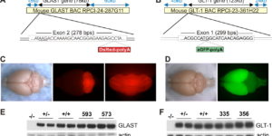
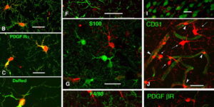
NG2 glia constitute a fourth major glial cell type in the mammalian central nervous system (CNS) that is distinct from other cell types. Although circumstantial evidence suggests that some NG2 glia differentiate into oligodendrocytes, their in vivo fate has not been directly examined. We have used the bacterial artificial chromosome (BAC) modification technique to generate transgenic mice that express DsRed or Cre specifically in NG2-expressing (NG2+) cells. In NG2DsRedBAC transgenic mice, DsRed was expressed specifically in NG2+ cells throughout the postnatal CNS. When the differentiation potential of NG2+ cells in vitro was examined using DsRed+NG2+ cells purified from perinatal transgenic brains, the majority of the cells either remained as NG2+ cells or differentiated into oligodendrocytes. In addition, DsRed+NG2+ cells also differentiated into astrocytes. The in vivo fate of NG2 glia was examined in mice that were double transgenic for NG2creBAC and the Cre reporter Z/EG. In the double transgenic mice, the Cre reporter EGFP was detected in myelinating oligodendrocytes and in a subpopulation of protoplasmic astrocytes in the gray matter of ventrolateral forebrain but not in fibrous astrocytes of white matter. These observations suggest that NG2+ cells are precursors of oligodendrocytes and some protoplasmic astrocytes in gray matter.

Variations in promoter activity reveal a differential expression and physiology of glutamate transporters by glia in the developing and mature CNS

Glutamate transporters regulate excitatory neurotransmission and prevent glutamate-mediated excitotoxicity in the CNS. To better study the cellular and temporal dynamics of the expression of these transporters, we generated bacterial artificial chromosome promoter Discosoma red [glutamate-aspartate transporter (GLAST)] and green fluorescent protein [glutamate transporter-1 (GLT-1)] reporter transgenic mice. Analysis of these mice revealed a differential activation of the transporter promoters not previously appreciated. GLT-1 promoter activity in the adult CNS is almost completely restricted to astrocytes, often and unexpectedly in a nonoverlapping pattern with GLAST. Spinal cord GLT-1 promoter reporter, protein density, and physiology were 10-fold lower than in brain, suggesting a possible mechanism for regional sensitivity seen in disease. The GLAST promoter is active in both radial glia and many astrocytes in the developing CNS but is downregulated in most astrocytes as the mice mature. In the adult CNS, the highest GLAST promoter activity was observed in radial glia, such as those located in the subgranular layer of the dentate gyrus. The continued expression of GLAST by these neural progenitors raises the possibility that GLAST may have an unanticipated role in regulating their behavior. In addition, GLAST promoter activation was observed in oligodendrocytes in white matter throughout many (e.g., spinal cord and corpus callosum), but not all (e.g., cerebellum), CNS fiber tracts. Overall, these studies of GLT-1 and GLAST promoter activity, protein expression, and glutamate uptake revealed a close correlation between transgenic reporter signals and uptake capacity, indicating that these mice provide the means to monitor the expression and regulation of glutamate transporters in situ.

Defining the role of astrocytes in neuromodulation

Astrocytes undergo elevations in intracellular calcium following activation of metabotropic receptors, which may trigger glutamate secretion and excitation of surrounding neurons. In this issue of Neuron, Fiacco et al. use transgenic mice that express a foreign G(q)-coupled receptor in astrocytes to show that selective stimulation of astrocytes is not sufficient to induce the release of glutamate.

Glutamate transporters bring competition to the synapse

Glutamate transporters (GluTs) prevent the accumulation of glutamate and influence the occupancy of receptors at synapses. The ability of extrasynaptic NMDA receptors and metabotropic glutamate receptors to participate in signaling is tightly regulated by GluT activity. Astrocytes express the highest density of GluTs and dominate clearance away from these receptors; synapses that are not associated with astrocyte processes experience greater mGluR activation and can be exposed to glutamate released at adjacent synapses. Although less abundant, neuronal transporters residing in the postsynaptic membrane can also shield receptors from the glutamate that is released. The diversity in synaptic morphology suggests a correspondingly rich diversity of GluT function in excitatory transmission.

Clearance of glutamate inside the synapse and beyond

The heated debate over the level of postsynaptic receptor occupancy by transmitter has not been extinguished – indeed, new evidence is fanning the flames. Recent experiments using two-photon microscopy suggest that the concentration of glutamate in the synaptic cleft does not attain levels previously suggested. In contrast, recordings from glial cells and studies of extrasynaptic receptor activation indicate that significant quantities of glutamate escape from the cleft following exocytosis. Determining the amount of glutamate efflux from the synaptic cleft and the distance it diffuses is critical to issues of synaptic specificity and the induction of synaptic plasticity.

Glial contribution to glutamate uptake at Schaffer collateral-commissural synapses in the hippocampus

Astrocytes in the hippocampus express high-affinity glutamate transporters that are important for lowering the concentration of extracellular glutamate after release at excitatory synapses. These transporters exhibit a permeability to chaotropic anions that is associated with transport, allowing their activity to be monitored in cell-fee patches when highly permeant anions are present. Astrocyte glutamate transporters are highly temperature sensitive, because L-glutamate-activated, anion-potentiated transporter currents in outside-out patches from these cells exhibited larger amplitudes and faster kinetics at 36 degreesC than at 24 degreesC. The cycling rate of these transporters was estimated by using paired applications of either L-glutamate or D-aspartate to measure the time necessary for the peak of the transporter current to recover from the steady-state level. Transporter currents in patches recovered with a time constant of 11.6 msec at 36 degreesC, suggesting that either the turnover rate of native transporters is much faster than previously reported for expressed EAAT2 transporters or the efficiency of these transporters is very low. Synaptically activated transporter currents persisted in astrocytes at physiological temperatures, although no evidence of these currents was found in CA1 pyramidal neurons in response to afferent stimulation. L-glutamate-gated transporter currents were also not detected in outside-out patches from pyramidal neurons. These results are consistent with the hypothesis that astrocyte transporters are responsible for taking up the majority of glutamate released at Schaffer collateral-commissural synapses in the hippocampus.Glial contribution to glutamate uptake at Schaffer collateral-commissural synapses in the hippocampus

Glutamate Release Monitored with Astrocyte Transporter Currents during LTP

Long-term potentiation (LTP) of synaptic transmission in the CA1 region of the hippocampus is thought to result from either increased transmitter release, heightened postsynaptic sensitivity, or a combination of the two. We have measured evoked glutamate release from Schaffer collateral/commissural fiber terminals in CA1 by recording synaptically activated glutamate transporter currents in hippocampal astrocytes located in stratum radiatum. Although several manipulations of release probability caused parallel changes in extracellular field potentials and synaptically activated transporter current amplitudes, induction of LTP failed to alter transporter-mediated responses, suggesting that LTP does not alter the amount of glutamate released upon synaptic stimulation.

Synaptic activation of glutamate transporters in hippocampal astrocytes

Glutamate transporters in the CNS are expressed in neurons and glia and mediate high affinity, electrogenic uptake of extracellular glutamate. Although glia have the highest capacity for glutamate uptake, the amount of glutamate that reaches glial membranes following release and the rate that glial transporters bind and sequester transmitter is not known. We find that stimulation of Schaffer collateral/commissural fibers in hippocampal slices evokes glutamate transporter currents in CA1 astrocytes that activate rapidly, indicating that a significant amount of transmitter escapes the synaptic cleft shortly after release. Transporter currents in outside-out patches from astrocytes have faster kinetics than synaptically elicited currents, suggesting that the glutamate concentration attained at astrocytic membranes is lower but remains elevated for longer than in the synaptic cleft.