Glutamate transporter currents in Bergmann glial cells follow the time course of extrasynaptic glutamate.

Glutamate transporters in the central nervous system are expressed in both neurons and glia, they mediate high affinity, electrogenic uptake of glutamate, and they are associated with an anion conductance that is stoichiometrically uncoupled from glutamate flux. Although a complete cycle of transport may require 50-100 ms, previous studies suggest that transporters can alter synaptic currents on a much faster time scale. We find that application of L-glutamate to outside-out patches from cerebellar Bergmann glia activates anion-potentiated glutamate transporter currents that activate in <1 ms, suggesting an efficient mechanism for the capture of extrasynaptic glutamate. Stimulation in the granule cell layer in cerebellar slices elicits all or none alpha-amino-3-hydroxy-5-methyl-4-isoxazolepropionate receptor and glutamate transporter currents in Bergmann glia that have a rapid onset, suggesting that glutamate released from climbing fiber terminals escapes synaptic clefts and reaches glial membranes shortly after release. Comparison of the concentration dependence of both alpha-amino-3-hydroxy-5-methyl-4-isoxazolepropionate receptor and glutamate transporter kinetics in patches with the time course of climbing fiber-evoked responses indicates that the glutamate transient at Bergmann glial membranes reaches a lower concentration than attained in the synaptic cleft and remains elevated in the extrasynaptic space for many milliseconds.
Synaptic activation of glutamate transporters in hippocampal astrocytes

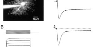
Glutamate transporters in the CNS are expressed in neurons and glia and mediate high affinity, electrogenic uptake of extracellular glutamate. Although glia have the highest capacity for glutamate uptake, the amount of glutamate that reaches glial membranes following release and the rate that glial transporters bind and sequester transmitter is not known. We find that stimulation of Schaffer collateral/commissural fibers in hippocampal slices evokes glutamate transporter currents in CA1 astrocytes that activate rapidly, indicating that a significant amount of transmitter escapes the synaptic cleft shortly after release. Transporter currents in outside-out patches from astrocytes have faster kinetics than synaptically elicited currents, suggesting that the glutamate concentration attained at astrocytic membranes is lower but remains elevated for longer than in the synaptic cleft.

Excitatory actions of norepinephrine on multiple classes of hippocampal CA1 interneurons

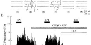
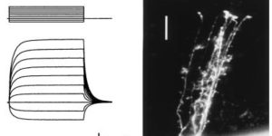
Norepinephrine (NE) causes an increase in the frequency of inhibitory postsynaptic potentials in CA1 pyramidal neurons in vitro. The possibility that this increase in tonic inhibition is caused by an excitatory effect on inhibitory interneurons was investigated through whole-cell recordings from pyramidal cells and both whole-cell and cell-attached patch recordings from visualized interneurons in acute slices of rat hippocampus. Adrenergic agonists caused a large increase in the frequency and amplitude of spontaneous IPSCs recorded from pyramidal cells in the presence of ionotropic glutamate receptor blockers, but they had no effect on either the frequency or the amplitude of action potential-independent miniature IPSCs recorded in tetrodotoxin. This effect was mediated primarily by an alpha adrenoceptor, although a slight beta adrenoceptor-dependent increase in IPSCs was also observed. NE caused interneurons located in all strata to depolarize and begin firing action potentials. Many of these cells had axons that ramified throughout the stratum pyramidale, suggesting that they are responsible for the IPSCs observed in pyramidal neurons. This depolarization was also mediated by an alpha adrenoceptor and was blocked by a selective alpha 1- but not a selective alpha 2-adrenoceptor antagonist. However, a slight beta adrenoceptor-dependent depolarization was detected in those interneurons that displayed time-dependent inward rectification. In the presence of a beta antagonist, NE induced an inward current that reversed near the predicted K+ equilibrium potential and was not affected by changes in intracellular Cl- concentration. In the presence of an alpha 1 antagonist, NE induced an inwardly rectifying current at potentials negative to approximately -70 mV that did not reverse (between -130 and -60 mV), characteristics similar to the hyperpolarization-activated current (lh). However, the depolarizing action of NE is attributable primarily to the alpha 1 adrenoceptor-mediated decrease in K+ conductance and not the beta adrenoceptor-dependent increase in lh. These results provide evidence that NE increases action potential-dependent IPSCs in pyramidal neurons by depolarizing surrounding inhibitory interneurons. This potent excitatory action of NE on multiple classes of hippocampal interneurons may contribute to the NE-induced decrease in the spontaneous activity of pyramidal neurons and the antiepileptic effects of NE observed in vivo.

Mossy fiber growth and synaptogenesis in rat hippocampal slices in vitro

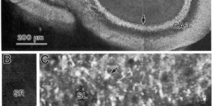
Hippocampal slices from early postnatal rat were used to study mossy fiber (MF) growth and synaptogenesis. The ability of MFs to form new giant synapses within isolated tissue slices was established by a series of experiments involving synapsin I immunohistochemistry, electron microscopy, and whole-cell recordings. When hippocampal slices from immature rats were cultured for up to 2 weeks, the distribution of giant MF terminals was similar to that found in vivo. Using a lesioning procedure, we determined that MFs in slices extend and form appropriate synaptic connections with normal target CA3 pyramidal cells. MF terminals were dispersed more widely than normal within the CA3 pyramidal layer after a lesion, but electron microscopy indicated that synaptic junctions were still primarily associated with pyramidal cell dendrites and not the somata. Establishment of functional synaptic input in vitro was confirmed by whole-cell recordings of MF-driven excitatory postsynaptic currents (50 pA to 1 nA) in pyramidal cells. The results establish for the first time that an MF projection with appropriate and functional synaptic connections can be formed de novo and not just maintained in excised hippocampal slices. The cellular dynamics underlying MF growth and synaptogenesis were examined directly by time-lapse confocal imaging of fibers selectively stained with a fluorescent membrane dye (Dil or DiO). MFs growing deep within isolated tissue slices were tipped by small (5-10 microns), active growth cones that advanced at variable rates (5-25 microns/hr). Furthermore, dynamic filopodial structures were seen at small varicosities along the length of developing MFs, which may identify nascent en passant synaptic contacts. The hippocampal slice preparations are shown to support normal development of MF connections and allow for direct visualization of the cellular dynamics of synapse formation in a mammalian CNS tissue environment.