A selective thyroid hormone β receptor agonist enhances human and rodent oligodendrocyte differentiation.

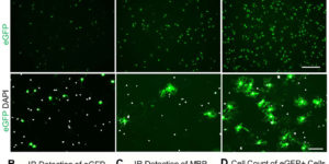
Nerve conduction within the mammalian central nervous system is made efficient by oligodendrocyte-derived myelin. Historically, thyroid hormones have a well described role in regulating oligodendrocyte differentiation and myelination during development; however, it remains unclear which thyroid hormone receptors are required to drive these effects. This is a question with clinical relevance since nonspecific thyroid receptor stimulation can produce deleterious side-effects. Here we report that GC-1, a thyromimetic with selective thyroid receptor β action and a potentially limited side-effect profile, promotes in vitro oligodendrogenesis from both rodent and human oligodendrocyte progenitor cells. In addition, we used in vivo genetic fate tracing of oligodendrocyte progenitor cells via PDGFαR-CreER;Rosa26-eYFP double-transgenic mice to examine the effect of GC-1 on cellular fate and find that treatment with GC-1 during developmental myelination promotes oligodendrogenesis within the corpus callosum, occipital cortex and optic nerve. GC-1 was also observed to enhance the expression of the myelin proteins MBP, CNP and MAG within the same regions. These results indicate that a β receptor selective thyromimetic can enhance oligodendrocyte differentiation in vitro and during developmental myelination in vivo and warrants further study as a therapeutic agent for demyelinating models.
Multiscale optical Ca2+ imaging of tonal organization in mouse auditory cortex.

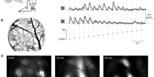
Spatial patterns of functional organization, resolved by microelectrode mapping, comprise a core principle of sensory cortices. In auditory cortex, however, recent two-photon Ca2+ imaging challenges this precept, as the traditional tonotopic arrangement appears weakly organized at the level of individual neurons. To resolve this fundamental ambiguity about the organization of auditory cortex, we developed multiscale optical Ca2+ imaging of unanesthetized GCaMP transgenic mice. Single-neuron activity monitored by two-photon imaging was precisely registered to large-scale cortical maps provided by transcranial widefield imaging. Neurons in the primary field responded well to tones; neighboring neurons were appreciably cotuned, and preferred frequencies adhered tightly to a tonotopic axis. By contrast, nearby secondary-field neurons exhibited heterogeneous tuning. The multiscale imaging approach also readily localized vocalization regions and neurons. Altogether, these findings cohere electrode and two-photon perspectives, resolve new features of auditory cortex, and offer a promising approach generalizable to any cortical area.

Norepinephrine controls astroglial responsiveness to local circuit activity.

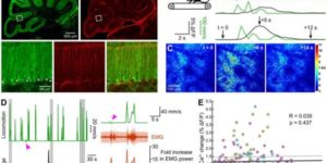
Astrocytes perform crucial supportive functions, including neurotransmitter clearance, ion buffering, and metabolite delivery. They can also influence blood flow and neuronal activity by releasing gliotransmitters in response to intracellular Ca(2+) transients. However, little is known about how astrocytes are engaged during different behaviors in vivo. Here we demonstrate that norepinephrine primes astrocytes to detect changes in cortical network activity. We show in mice that locomotion triggers simultaneous activation of astrocyte networks in multiple brain regions. This global stimulation of astrocytes was inhibited by alpha-adrenoceptor antagonists and abolished by depletion of norepinephrine from the brain. Although astrocytes in visual cortex of awake mice were rarely engaged when neurons were activated by light stimulation alone, pairing norepinephrine release with light stimulation markedly enhanced astrocyte Ca(2+) signaling. Our findings indicate that norepinephrine shifts the gain of astrocyte networks according to behavioral state, enabling astrocytes to respond to local changes in neuronal activity.

The blood-brain barrier: an engineering perspective.

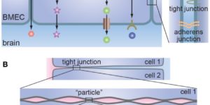
It has been more than 100 years since Paul Ehrlich reported that various water-soluble dyes injected into the circulation did not enter the brain. Since Ehrlich’s first experiments, only a small number of molecules, such as alcohol and caffeine have been found to cross the blood-brain barrier, and this selective permeability remains the major roadblock to treatment of many central nervous system diseases. At the same time, many central nervous system diseases are associated with disruption of the blood-brain barrier that can lead to changes in permeability, modulation of immune cell transport, and trafficking of pathogens into the brain. Therefore, advances in our understanding of the structure and function of the blood-brain barrier are key to developing effective treatments for a wide range of central nervous system diseases. Over the past 10 years it has become recognized that the blood-brain barrier is a complex, dynamic system that involves biomechanical and biochemical signaling between the vascular system and the brain. Here we reconstruct the structure, function, and transport properties of the blood-brain barrier from an engineering perspective. New insight into the physics of the blood-brain barrier could ultimately lead to clinical advances in the treatment of central nervous system diseases.

Oligodendrocyte progenitors balance growth with self-repulsion to achieve homeostasis in the adult brain.

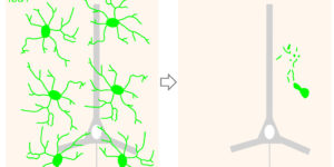
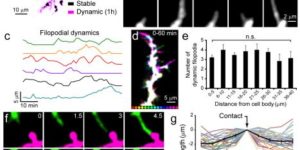
The adult CNS contains an abundant population of oligodendrocyte precursor cells (NG2(+) cells) that generate oligodendrocytes and repair myelin, but how these ubiquitous progenitors maintain their density is unknown. We generated NG2-mEGFP mice and used in vivo two-photon imaging to study their dynamics in the adult brain. Time-lapse imaging revealed that NG2(+) cells in the cortex were highly dynamic; they surveyed their local environment with motile filopodia, extended growth cones and continuously migrated. They maintained unique territories though self-avoidance, and NG2(+) cell loss though death, differentiation or ablation triggered rapid migration and proliferation of adjacent cells to restore their density. NG2(+) cells recruited to sites of focal CNS injury were similarly replaced by a proliferative burst surrounding the injury site. Thus, homeostatic control of NG2(+) cell density through a balance of active growth and self-repulsion ensures that these progenitors are available to replace oligodendrocytes and participate in tissue repair.

Degeneration and impaired regeneration of gray matter oligodendrocytes in amyotrophic lateral sclerosis.

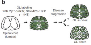
Oligodendrocytes associate with axons to establish myelin and provide metabolic support to neurons. In the spinal cord of amyotrophic lateral sclerosis (ALS) mice, oligodendrocytes downregulate transporters that transfer glycolytic substrates to neurons and oligodendrocyte progenitors (NG2(+) cells) exhibit enhanced proliferation and differentiation, although the cause of these changes in oligodendroglia is unknown. We found extensive degeneration of gray matter oligodendrocytes in the spinal cord of SOD1 (G93A) ALS mice prior to disease onset. Although new oligodendrocytes were formed, they failed to mature, resulting in progressive demyelination. Oligodendrocyte dysfunction was also prevalent in human ALS, as gray matter demyelination and reactive changes in NG2(+) cells were observed in motor cortex and spinal cord of ALS patients. Selective removal of mutant SOD1 from oligodendroglia substantially delayed disease onset and prolonged survival in ALS mice, suggesting that ALS-linked genes enhance the vulnerability of motor neurons and accelerate disease by directly impairing the function of oligodendrocytes.

Reduction of motion artifacts during in vivo two-photon imaging of brain through heartbeat triggered scanning.

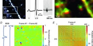
Two-photon imaging of fluorescence in brain enables analysis of the structure and dynamic activity of neurons and glial cells in living animals. However, vital functions such as beating of the heart cause pulsations in brain tissue, leading to image distortion and loss of resolution. We find that synchronizing imaging scans to the cardiac cycle reduces motion artifacts, significantly improving the resolution of cellular structures. By interlacing multiple heartbeat triggered imaging scans, it was possible to image large brain volumes with negligible distortion. This approach can be readily incorporated into conventional microscopes to achieve substantial reductions in motion artifacts during two-photon imaging.

The density of EAAC1 (EAAT3) glutamate transporters expressed by neurons in the mammalian CNS.

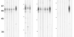
The extracellular levels of excitatory amino acids are kept low by the action of the glutamate transporters. Glutamate/aspartate transporter (GLAST) and glutamate transporter-1 (GLT-1) are the most abundant subtypes and are essential for the functioning of the mammalian CNS, but the contribution of the EAAC1 subtype in the clearance of synaptic glutamate has remained controversial, because the density of this transporter in different tissues has not been determined. We used purified EAAC1 protein as a standard during immunoblotting to measure the concentration of EAAC1 in different CNS regions. The highest EAAC1 levels were found in the young adult rat hippocampus. Here, the concentration of EAAC1 was ∼0.013 mg/g tissue (∼130 molecules μm⁻³), 100 times lower than that of GLT-1. Unlike GLT-1 expression, which increases in parallel with circuit formation, only minor changes in the concentration of EAAC1 were observed from E18 to adulthood. In hippocampal slices, photolysis of MNI-D-aspartate (4-methoxy-7-nitroindolinyl-D-aspartate) failed to elicit EAAC1-mediated transporter currents in CA1 pyramidal neurons, and D-aspartate uptake was not detected electron microscopically in spines. Using EAAC1 knock-out mice as negative controls to establish antibody specificity, we show that these relatively small amounts of EAAC1 protein are widely distributed in somata and dendrites of all hippocampal neurons. These findings raise new questions about how so few transporters can influence the activation of NMDA receptors at excitatory synapses.