NgR1 and NgR3 are receptors for chondroitin sulfate proteoglycans.

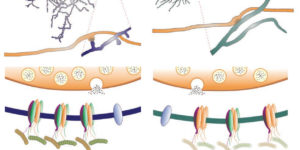
In the adult mammalian CNS, chondroitin sulfate proteoglycans (CSPGs) and myelin-associated inhibitors (MAIs) stabilize neuronal structure and restrict compensatory sprouting following injury. The Nogo receptor family members NgR1 and NgR2 bind to MAIs and have been implicated in neuronal inhibition. We found that NgR1 and NgR3 bind with high affinity to the glycosaminoglycan moiety of proteoglycans and participate in CSPG inhibition in cultured neurons. Nogo receptor triple mutants (Ngr1(-/-); Ngr2(-/-); Ngr3(-/-); which are also known as Rtn4r, Rtn4rl2 and Rtn4rl1, respectively), but not single mutants, showed enhanced axonal regeneration following retro-orbital optic nerve crush injury. The combined loss of Ngr1 and Ngr3 (Ngr1(-/-); Ngr3(-/-)), but not Ngr1 and Ngr2 (Ngr1(-/-); Ngr2(-/-)), was sufficient to mimic the triple mutant regeneration phenotype. Regeneration in Ngr1(-/-); Ngr3(-/-) mice was further enhanced by simultaneous ablation of Rptpσ (also known as Ptprs), a known CSPG receptor. Collectively, our results identify NgR1 and NgR3 as CSPG receptors, suggest that there is functional redundancy among CSPG receptors, and provide evidence for shared mechanisms of MAI and CSPG inhibition.
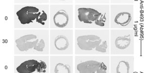
Specificity controls for immunocytochemistry: the antigen preadsorption test can lead to inaccurate assessment of antibody specificity.

The biomedical research community relies directly or indirectly on immunocytochemical data. Unfortunately, validation of labeling specificity is difficult. A common specificity test is the preadsorption test. This test was intended for testing crude antisera but is now frequently used to validate monoclonal and affinity purified polyclonal antibodies. Here, the authors assess the power of this test. Nine affinity purified antibodies to different epitopes on 3 proteins (EAAT3, slc1a1; EAAT2, slc1a2; BGT1, slc6a12) were tested on samples (tissue sections and Western blots with or without fixation). The selected antibodies displayed some degree of cross-reactivity as defined by labeling of samples from knockout mice. The authors show that antigen preadsorption blocked all labeling of both wild-type and knockout samples, implying that preadsorption also blocked binding to cross-reactive epitopes. They show how this can give an illusion of specificity and illustrate sensitivity-specificity relationships, the importance of good negative controls, that fixation can create new epitopes, and that cross-reacting epitopes present in sections may not be present on Western blots and vice versa. In conclusion, they argue against uncritical use of the preadsorption test and, in doing so, address a number of other issues related to immunocytochemistry specificity testing.

Neuronal activity regulates glutamate transporter dynamics in developing astrocytes.

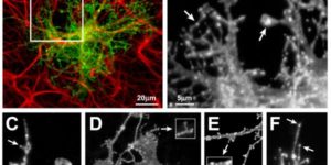
Glutamate transporters (GluTs) maintain a low ambient level of glutamate in the central nervous system (CNS) and shape the activation of glutamate receptors at synapses. Nevertheless, the mechanisms that regulate the trafficking and localization of transporters near sites of glutamate release are poorly understood. Here, we examined the subcellular distribution and dynamic remodeling of the predominant GluT GLT-1 (excitatory amino acid transporter 2, EAAT2) in developing hippocampal astrocytes. Immunolabeling revealed that endogenous GLT-1 is concentrated into discrete clusters along branches of developing astrocytes that were apposed preferentially to synapsin-1 positive synapses. Green fluorescent protein (GFP)-GLT-1 fusion proteins expressed in astrocytes also formed distinct clusters that lined the edges of astrocyte processes, as well as the tips of filopodia and spine-like structures. Time-lapse three-dimensional confocal imaging in tissue slices revealed that GFP-GLT-1 clusters were dynamically remodeled on a timescale of minutes. Some transporter clusters moved within developing astrocyte branches as filopodia extended and retracted, while others maintained stable positions at the tips of spine-like structures. Blockade of neuronalactivity with tetrodotoxin reduced both the density and perisynaptic localization of GLT-1 clusters. Conversely, enhancement of neuronal activity increased the size of GLT-1 clusters and their proximity to synapses. Together, these findings indicate that neuronal activity influences both the organization of GluTs in developing astrocyte membranes and their position relative to synapses.

NMDA receptor signaling in oligodendrocyte progenitors is not required for oligodendrogenesis and myelination.

Oligodendrocyte precursor cells (OPCs) express NMDA receptors (NMDARs) and form synapses with glutamatergic neurons throughout the CNS. Although glutamate influences the proliferation and maturation of these progenitors in vitro, the role of NMDAR signaling in oligodendrogenesis and myelination in vivo is not known. Here, we investigated the consequences of genetically deleting the obligatory NMDAR subunit NR1 from OPCs and their oligodendrocyte progeny in the CNS of developing and mature mice. NMDAR-deficient OPCs proliferated normally, achieved appropriate densities in gray and white matter, and differentiated to form major white matter tracts without delay. OPCs also retained their characteristic physiological and morphological properties in the absence of NMDAR signaling and were able to form synapses with glutamatergic axons. However, expression of calcium-permeable AMPA receptors (AMPARs) was enhanced in NMDAR-deficient OPCs. These results suggest that NMDAR signaling is not used to control OPC development but to regulate AMPAR-dependent signaling with surrounding axons, pointing to additional functions for these ubiquitous glial cells.

A requirement for nuclear factor-kappaB in developmental and plasticity-associated synaptogenesis.

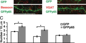
Structural plasticity of dendritic spines and synapses is a fundamental mechanism governing neuronal circuits and may form an enduring basis for information storage in the brain. We find that the p65 subunit of the nuclear factor-κB (NF-κB) transcription factor, which is required for learning and memory, controls excitatory synapse and dendritic spine formation and morphology in murine hippocampal neurons. Endogenous NF-κB activity is elevated by excitatory transmission during periods of rapid spine and synapse development. During in vitro synaptogenesis, NF-κB enhances dendritic spine and excitatory synapse density and loss of endogenous p65 decreases spine density and spine head volume. Cell-autonomous function of NF-κB within the postsynaptic neuron is sufficient to regulate the formation of both presynaptic and postsynaptic elements. During synapse development in vivo, loss of NF-κB similarly reduces spine density and also diminishes the amplitude of synaptic responses. In contrast, after developmental synaptogenesis has plateaued, endogenous NF-κB activity is low and p65 deficiency no longer attenuates basal spine density. Instead, NF-κB in mature neurons is activated by stimuli that induce demand for new synapses, including estrogen and short-term bicuculline, and is essential for upregulating spine density in response to these stimuli. p65 is enriched in dendritic spines making local protein-protein interactions possible; however, the effects of NF-κB on spine density require transcription and the NF-κB-dependent regulation of PSD-95, a critical postsynaptic component. Collectively, our data define a distinct role for NF-κB in imparting transcriptional regulation required for the induction of changes to, but not maintenance of, excitatory synapse and spine density.

NG2+ CNS glial progenitors remain committed to the oligodendrocyte lineage in postnatal life and following neurodegeneration.

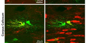
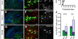
The mammalian CNS contains a ubiquitous population of glial progenitors known as NG2+ cells that have the ability to develop into oligodendrocytes and undergo dramatic changes in response to injury and demyelination. Although it has been reported that NG2+ cells are multipotent, their fate in health and disease remains controversial. Here, we generated PDGFαR-CreER transgenic mice and followed their fate in vivo in the developing and adult CNS. These studies revealed that NG2+ cells in the postnatal CNS generate myelinating oligodendrocytes, but not astrocytes or neurons. In regions of neurodegeneration in the spinal cord of ALS mice, NG2+ cells exhibited enhanced proliferation and accelerated differentiation into oligodendrocytes but remained committed to the oligodendrocyte lineage. These results indicate that NG2+ cells in the normal CNS are oligodendrocyte precursors with restricted lineage potential and that cell loss and gliosis are not sufficient to alter the lineage potential of these progenitors.

Calcium action potentials in hair cells pattern auditory neuron activity before hearing onset.

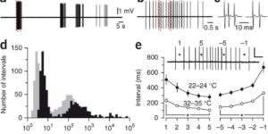
We found rat central auditory neurons to fire action potentials in a precise sequence of mini-bursts before the age of hearing onset. This stereotyped pattern was initiated by hair cells in the cochlea, which trigger brief bursts of action potentials in auditory neurons each time they fire a Ca2+ spike. By generating theta-like activity, hair cells may limit the influence of synaptic depression in developing auditory circuits and promote consolidation of synapses.

ATP-induced morphological changes in supporting cells of the developing cochlea.

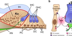
The developing cochlea of mammals contains a large group of columnar-shaped cells, which together form a structure known as Kölliker’s organ. Prior to the onset of hearing, these inner supporting cells periodically release adenosine 5′-triphosphate (ATP), which activates purinergic receptors in surrounding supporting cells, inner hair cells and the dendrites of primary auditory neurons. Recent studies indicate that purinergic signaling between inner supporting cells and inner hair cells initiates bursts of action potentials in auditory nerve fibers before the onset of hearing. ATP also induces prominent effects in inner supporting cells, including an increase in membrane conductance, a rise in intracellular Ca(2+), and dramatic changes in cell shape, although the importance of ATP signaling in non-sensory cells of the developing cochlea remains unknown. Here, we review current knowledge pertaining to purinergic signaling in supporting cells of Kölliker’s organ and focus on the mechanisms by which ATP induces changes in their morphology. We show that these changes in cell shape are preceded by increases in cytoplasmic Ca(2+), and provide new evidence indicating that elevation of intracellular Ca(2+) and IP(3) are sufficient to initiate shape changes. In addition, we discuss the possibility that these ATP-mediated morphological changes reflect crenation following the activation of Ca(2+)-activated Cl(-) channels, and speculate about the possible functions of these changes in cell morphology for maturation of the cochlea.

Zones of enhanced glutamate release from climbing fibers in the mammalian cerebellum.

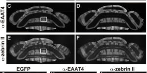
Purkinje cells in the mammalian cerebellum are remarkably homogeneous in shape and orientation, yet they exhibit regional differences in gene expression. Purkinje cells that express high levels of zebrin II (aldolase C) and the glutamate transporter EAAT4 cluster in parasagittal zones that receive input from distinct groups of climbing fibers (CFs); however, the physiological properties of CFs that target these molecularly distinct Purkinje cells have not been determined. Here we report that CFs that innervate Purkinje cells in zebrin II-immunoreactive (Z(+)) zones release more glutamate per action potential than CFs in Z(-) zones. CF terminals in Z(+) zones had larger pools of release-ready vesicles, exhibited enhanced multivesicular release, and produced larger synaptic glutamate transients. As a result, CF-mediated EPSCs in Purkinje cells decayed more slowly in Z(+) zones, which triggered longer-duration complex spikes containing a greater number of spikelets. The differences in the duration of CF EPSCs between Z(+) and Z(-) zones persisted in EAAT4 knock-out mice, indicating that EAAT4 is not required for maintaining this aspect of CF function. These results indicate that the organization of the cerebellum into discrete longitudinal zones is defined not only by molecular phenotype of Purkinje cells within zones, but also by the physiological properties of CFs that project to these distinct regions. The enhanced release of glutamate from CFs in Z(+) zones may alter the threshold for synaptic plasticity and prolong inhibition of cerebellar output neurons in deep cerebellar nuclei.