Coupled Activation of Primary Sensory Neurons Contributes to Chronic Pain.

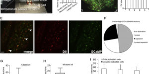
Primary sensory neurons in the DRG play an essential role in initiating pain by detecting painful stimuli in the periphery. Tissue injury can sensitize DRG neurons, causing heightened pain sensitivity, often leading to chronic pain. Despite the functional importance, how DRG neurons function at a population level is unclear due to the lack of suitable tools. Here we developed an imaging technique that allowed us to simultaneously monitor the activities of >1,600 neurons/DRG in live mice and discovered a striking neuronal coupling phenomenon that adjacent neurons tend to activate together following tissue injury. This coupled activation occurs among various neurons and is mediated by an injury-induced upregulation of gap junctions in glial cells surrounding DRG neurons. Blocking gap junctions attenuated neuronal coupling and mechanical hyperalgesia. Therefore, neuronal coupling represents a new form of neuronal plasticity in the DRG and contributes to pain hypersensitivity by “hijacking” neighboring neurons through gap junctions.

NMDA Receptors Enhance Spontaneous Activity and Promote Neuronal Survival in the Developing Cochlea.

Spontaneous bursts of activity in developing sensory pathways promote maturation of neurons, refinement of neuronal connections, and assembly of appropriate functional networks. In the developing auditory system, inner hair cells (IHCs) spontaneously fire Ca(2+) spikes, each of which is transformed into a mini-burst of action potentials in spiral ganglion neurons (SGNs). Here we show that NMDARs are expressed in SGN dendritic terminals and play a critical role during transmission of activity from IHCs to SGNs before hearing onset. NMDAR activation enhances glutamate-mediated Ca(2+) influx at dendritic terminals, promotes repetitive firing of individual SGNs in response to each synaptic event, and enhances coincident activity of neighboring SGNs that will eventually encode similar frequencies of sound. Loss of NMDAR signaling from SGNs reduced their survival both in vivo and in vitro, revealing that spontaneous activity in the prehearing cochlea promotes maturation of auditory circuitry through periodic activation of NMDARs in SGNs.

Spontaneous Activity of Cochlear Hair Cells Triggered by Fluid Secretion Mechanism in Adjacent Support Cells.

Spontaneous electrical activity of neurons in developing sensory systems promotes their maturation and proper connectivity. In the auditory system, spontaneous activity of cochlear inner hair cells (IHCs) is initiated by the release of ATP from glia-like inner supporting cells (ISCs), facilitating maturation of central pathways before hearing onset. Here, we find that ATP stimulates purinergic autoreceptors in ISCs, triggering Cl(-) efflux and osmotic cell shrinkage by opening TMEM16A Ca(2+)-activated Cl(-) channels. Release of Cl(-) from ISCs also forces K(+) efflux, causing transient depolarization of IHCs near ATP release sites. Genetic deletion of TMEM16A markedly reduces the spontaneous activity of IHCs and spiral ganglion neurons in the developing cochlea and prevents ATP-dependent shrinkage of supporting cells. These results indicate that supporting cells in the developing cochlea have adapted a pathway used for fluid secretion in other organs to induce periodic excitation of hair cells.

Neuron-glia Signaling in Developing Retina Mediated by Neurotransmitter Spillover.

Neuron-glia interactions play a critical role in the maturation of neural circuits; however, little is known about the pathways that mediate their communication in the developing CNS. We investigated neuron-glia signaling in the developing retina, where we demonstrate that retinal waves reliably induce calcium transients in Müller glial cells (MCs). During cholinergic waves, MC calcium transients were blocked by muscarinic acetylcholine receptor antagonists, whereas during glutamatergic waves, MC calcium transients were inhibited by ionotropic glutamate receptor antagonists, indicating that the responsiveness of MCs changes to match the neurotransmitter used to support retinal waves. Using an optical glutamate sensor we show that the decline in MC calcium transients is caused by a reduction in the amount of glutamate reaching MCs. Together, these studies indicate that neurons and MCs exhibit correlated activity during a critical period of retinal maturation that is enabled by neurotransmitter spillover from retinal synapses.

Spontaneous activity in the developing auditory system.

Spontaneous electrical activity is a common feature of sensory systems during early development. This sensory-independent neuronal activity has been implicated in promoting their survival and maturation, as well as growth and refinement of their projections to yield circuits that can rapidly extract information about the external world. Periodic bursts of action potentials occur in auditory neurons of mammals before hearing onset. This activity is induced by inner hair cells (IHCs) within the developing cochlea, which establish functional connections with spiral ganglion neurons (SGNs) several weeks before they are capable of detecting external sounds. During this pre-hearing period, IHCs fire periodic bursts of Ca(2+) action potentials that excite SGNs, triggering brief but intense periods of activity that pass through auditory centers of the brain. Although spontaneous activity requires input from IHCs, there is ongoing debate about whether IHCs are intrinsically active and their firing periodically interrupted by external inhibitory input (IHC-inhibition model), or are intrinsically silent and their firing periodically promoted by an external excitatory stimulus (IHC-excitation model). There is accumulating evidence that inner supporting cells in Kölliker’s organ spontaneously release ATP during this time, which can induce bursts of Ca(2+) spikes in IHCs that recapitulate many features of auditory neuron activity observed in vivo. Nevertheless, the role of supporting cells in this process remains to be established in vivo. A greater understanding of the molecular mechanisms responsible for generating IHC activity in the developing cochlea will help reveal how these events contribute to the maturation of nascent auditory circuits.

Early white matter abnormalities, progressive brain pathology and motor deficits in a novel knock-in mouse model of Huntington’s disease.

White matter abnormalities have been reported in premanifest Huntington’s disease (HD) subjects before overt striatal neuronal loss, but whether the white matter changes represent a necessary step towards further pathology and the underlying mechanism of these changes remains unknown. Here, we characterized a novel knock-in mouse model that expresses mouse HD gene homolog (Hdh) with extended CAG repeat- HdhQ250, which was derived from the selective breeding of HdhQ150 mice. HdhQ250 mice manifest an accelerated and robust phenotype compared with its parent line. HdhQ250 mice exhibit progressive motor deficits, reduction in striatal and cortical volume, accumulation of mutant huntingtin aggregation, decreased levels of DARPP32 and BDNF and altered striatal metabolites. The abnormalities detected in this mouse model are reminiscent of several aspects of human HD. In addition, disturbed myelination was evident in postnatal Day 14 HdhQ250 mouse brain, including reduced levels of myelin regulatory factor and myelin basic protein, and decreased numbers of myelinated axons in the corpus callosum. Thinner myelin sheaths, indicated by increased G-ratio of myelin, were also detected in the corpus callosum of adult HdhQ250 mice. Moreover, proliferation of oligodendrocyte precursor cells is altered by mutant huntingtin both in vitro and in vivo. Our data indicate that this model is suitable for understanding comprehensive pathogenesis of HD in white matter and gray matter as well as developing therapeutics for HD.

Human astrocytes develop physiological morphology and remain quiescent in a novel 3D matrix.

Astrocytes are the most abundant glial cells in the brain and are responsible for diverse functions, from modulating synapse function to regulating the blood-brain barrier. In vivo, these cells exhibit a star-shaped morphology with multiple radial processes that contact synapses and completely surround brain capillaries. In response to trauma or CNS disease, astrocytes become activated, a state associated with profound changes in gene expression, including upregulation of intermediate filament proteins, such as glial fibrillary acidic protein (GFAP). The inability to recapitulate the complex structure of astrocytes and maintain their quiescent state in vitro is a major roadblock to further developments in tissue engineering and regenerative medicine. Here, we characterize astrocyte morphology and activation in various hydrogels to assess the feasibility of developing a matrix that mimics key aspects of the native microenvironment. We show that astrocytes seeded in optimized matrix composed of collagen, hyaluronic acid, and matrigel exhibit a star-shaped morphology with radial processes and do not upregulate GFAP expression, hallmarks of quiescent astrocytes in the brain. In these optimized gels, collagen I provides structural support, HA mimics the brain extracellular matrix, and matrigel provides endothelial cell compatibility and was found to minimize GFAP upregulation. This defined 3D microenvironment for maintaining human astrocytes in vitro provides new opportunities for developing improved models of the blood-brain barrier and studying their response to stress signals.

Calcium dynamics in astrocyte processes during neurovascular coupling.

Enhanced neuronal activity in the brain triggers a local increase in blood flow, termed functional hyperemia, via several mechanisms, including calcium (Ca(2+)) signaling in astrocytes. However, recent in vivo studies have questioned the role of astrocytes in functional hyperemia because of the slow and sparse dynamics of their somatic Ca(2+) signals and the absence of glutamate metabotropic receptor 5 in adults. Here, we reexamined their role in neurovascular coupling by selectively expressing a genetically encoded Ca(2+) sensor in astrocytes of the olfactory bulb. We show that in anesthetized mice, the physiological activation of olfactory sensory neuron (OSN) terminals reliably triggers Ca(2+) increases in astrocyte processes but not in somata. These Ca(2+) increases systematically precede the onset of functional hyperemia by 1-2 s, reestablishing astrocytes as potential regulators of neurovascular coupling.

Entrapment via synaptic-like connections between NG2 proteoglycan+ cells and dystrophic axons in the lesion plays a role in regeneration failure after spinal cord injury.

NG2 is purportedly one of the most growth-inhibitory chondroitin sulfate proteoglycans (CSPGs) produced after spinal cord injury. Nonetheless, once the severed axon tips dieback from the lesion core into the penumbra they closely associate with NG2+ cells. We asked if proteoglycans play a role in this tight cell-cell interaction and whether overadhesion upon these cells might participate in regeneration failure in rodents. Studies using varying ratios of CSPGs and adhesion molecules along with chondroitinase ABC, as well as purified adult cord-derived NG2 glia, demonstrate that CSPGs are involved in entrapping neurons. Once dystrophic axons become stabilized upon NG2+ cells, they form synaptic-like connections both in vitro and in vivo. In NG2 knock-out mice, sensory axons in the dorsal columns dieback further than their control counterparts. When axons are double conditioned to enhance their growth potential, some traverse the lesion core and express reduced amounts of synaptic proteins. Our studies suggest that proteoglycan-mediated entrapment upon NG2+ cells is an additional obstacle to CNS axon regeneration.

Spontaneous regeneration of cochlear supporting cells after neonatal ablation ensures hearing in the adult mouse.

Supporting cells in the cochlea play critical roles in the development, maintenance, and function of sensory hair cells and auditory neurons. Although the loss of hair cells or auditory neurons results in sensorineural hearing loss, the consequence of supporting cell loss on auditory function is largely unknown. In this study, we specifically ablated inner border cells (IBCs) and inner phalangeal cells (IPhCs), the two types of supporting cells surrounding inner hair cells (IHCs) in mice in vivo. We demonstrate that the organ of Corti has the intrinsic capacity to replenish IBCs/IPhCs effectively during early postnatal development. Repopulation depends on the presence of hair cells and cells within the greater epithelial ridge and is independent of cell proliferation. This plastic response in the neonatal cochlea preserves neuronal survival, afferent innervation, and hearing sensitivity in adult mice. In contrast, the capacity for IBC/IPhC regeneration is lost in the mature organ of Corti, and consequently IHC survival and hearing sensitivity are impaired significantly, demonstrating that there is a critical period for the regeneration of cochlear supporting cells. Our findings indicate that the quiescent neonatal organ of Corti can replenish specific supporting cells completely after loss in vivo to guarantee mature hearing function.