ATP-induced morphological changes in supporting cells of the developing cochlea.
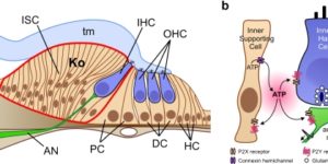
The developing cochlea of mammals contains a large group of columnar-shaped cells, which together form a structure known as Kölliker’s organ. Prior to the onset of hearing, these inner supporting cells periodically release adenosine 5′-triphosphate (ATP), which activates purinergic receptors in surrounding supporting cells, inner hair cells and the dendrites of primary auditory neurons. Recent studies indicate that purinergic signaling between inner supporting cells and inner hair cells initiates bursts of action potentials in auditory nerve fibers before the onset of hearing. ATP also induces prominent effects in inner supporting cells, including an increase in membrane conductance, a rise in intracellular Ca(2+), and dramatic changes in cell shape, although the importance of ATP signaling in non-sensory cells of the developing cochlea remains unknown. Here, we review current knowledge pertaining to purinergic signaling in supporting cells of Kölliker’s organ and focus on the mechanisms by which ATP induces changes in their morphology. We show that these changes in cell shape are preceded by increases in cytoplasmic Ca(2+), and provide new evidence indicating that elevation of intracellular Ca(2+) and IP(3) are sufficient to initiate shape changes. In addition, we discuss the possibility that these ATP-mediated morphological changes reflect crenation following the activation of Ca(2+)-activated Cl(-) channels, and speculate about the possible functions of these changes in cell morphology for maturation of the cochlea.