Remyelination Alters the Pattern of Myelin in the Cerebral Cortex

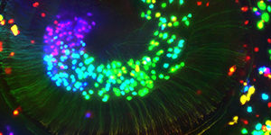
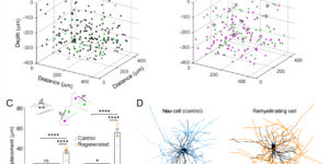
Destruction of oligodendrocytes and myelin sheaths in cortical gray matter profoundly alters neural activity and is associated with cognitive disability in multiple sclerosis (MS). Myelin can be restored by regenerating oligodendrocytes from resident progenitors; however, it is not known whether regeneration restores the complex myelination patterns in cortical circuits. Here we performed time lapse in vivo two photon imaging in somatosensory cortex of adult mice to define the kinetics and specificity of myelin regeneration after acute oligodendrocyte ablation. These longitudinal studies revealed that the pattern of myelination in cortex changed dramatically after regeneration, as new oligodendrocytes were formed in different locations and new sheaths were often established along axon segments previously lacking myelin. Despite the dramatic increase in axonal territory available, oligodendrogenesis was persistently impaired in deeper cortical layers that experienced higher gliosis. Repeated reorganization of myelin patterns in MS may alter circuit function and contribute to cognitive decline.

Changes in the Oligodendrocyte Progenitor Cell Proteome With Ageing

Following central nervous system (CNS) demyelination, adult oligodendrocyte progenitor cells (OPCs) can differentiate into new myelin-forming oligodendrocytes in a regenerative process called remyelination. While remyelination is very efficient in young adults, its efficiency declines progressively with ageing. Here we performed proteomic analysis of OPCs freshly isolated from the brains of neonate, young and aged female rats. Approximately 50% of the proteins are expressed at different levels in OPCs from neonates compared to their adult counterparts. The amount of myelin-associated proteins, and proteins associated with oxidative phosphorylation, inflammatory responses and actin cytoskeletal organization increased with age, while cholesterol-biosynthesis, transcription factors and cell cycle proteins decreased. Our experiments provide the first ageing OPC proteome, revealing the distinct features of OPCs at different ages. These studies provide new insights into why remyelination efficiency declines with ageing and potential roles for aged OPCs in other neurodegenerative diseases.

Persistent Cyfip1 Expression Is Required to Maintain the Adult Subventricular Zone Neurogenic Niche

Neural stem cells (NSCs) persist throughout life in the subventricular zone (SVZ) neurogenic niche of the lateral ventricles as Type B1 cells in adult mice. Maintaining this population of NSCs depends on the balance between quiescence and self-renewing or self-depleting cell divisions. Interactions between B1 cells and the surrounding niche are important in regulating this balance, but the mechanisms governing these processes have not been fully elucidated. The cytoplasmic FMRP-interacting protein (Cyfip1) regulates apical-basal polarity in the embryonic brain. Loss of Cyfip1 during embryonic development in mice disrupts the embryonic niche and affects cortical neurogenesis. However, a direct role for Cyfip1 in the regulation of adult NSCs has not been established. Here, we demonstrate that Cyfip1 expression is preferentially localized to B1 cells in the adult mouse SVZ. Loss of Cyfip1 in the embryonic mouse brain results in altered adult SVZ architecture and expansion of the adult B1 cell population at the ventricular surface. Furthermore, acute deletion of Cyfip1 in adult NSCs results in a rapid change in adherens junction proteins as well as increased proliferation and number of B1 cells at the ventricular surface. Together, these data indicate that Cyfip1 plays a critical role in the formation and maintenance of the adult SVZ niche; furthermore, deletion of Cyfip1 unleashes the capacity of adult B1 cells for symmetric renewal to increase the adult NSC pool.

Immune Cell Modulation of Oligodendrocyte Lineage Cells

Chronic demyelination and the concomitant loss of trophic support and increased energy demands in axons are thought to contribute to neurodegeneration in a number of neurological diseases such as multiple sclerosis (MS). Adult oligodendrocyte precursor cells (OPCs) play an important role in these demyelinating diseases by generating new myelinating oligodendrocytes that may help limit axonal degeneration. Thus, promoting the differentiation of OPCs and functional integration of newly generated oligodendrocytes is a crucial avenue for the next generation of therapies. Evidence to date suggests that the immune system may both positively and negatively impact OPC differentiation and endogenous remyelination in disease. Inflammatory cytokines not only suppress OPC differentiation but may also directly affect other functions of OPCs. Recent studies have demonstrated that OPCs and oligodendrocytes in both human multiple sclerosis lesions and mouse models of demyelination can express an immunogenic transcriptional signature and upregulate antigen presenting genes. In inflammatory demyelinating mouse models OPCs are capable of presenting antigen and activating CD8 + T cells. Here we review the evidence for this new role of oligodendroglia as antigen presenting cells and how these inflammatory OPCs (iOPCs) and inflammatory oligodendrocytes (iOLs) may influence myelin repair and other disease processes.

Purinergic Signaling in Cochlear Supporting Cells Reduces Hair Cell Excitability by Increasing the Extracellular Space

Neurons in developing sensory pathways exhibit spontaneous bursts of electrical activity that are critical for survival, maturation and circuit refinement. In the auditory system, intrinsically generated activity arises within the cochlea, but the molecular mechanisms that initiate this activity remain poorly understood. We show that burst firing of mouse inner hair cells prior to hearing onset requires P2RY1 autoreceptors expressed by inner supporting cells. P2RY1 activation triggers K+ efflux and depolarization of hair cells, as well as osmotic shrinkage of supporting cells that dramatically increased the extracellular space and speed of K+ redistribution. Pharmacological inhibition or genetic disruption of P2RY1 suppressed neuronal burst firing by reducing K+ release, but unexpectedly enhanced their tonic firing, as water resorption by supporting cells reduced the extracellular space, leading to K+ accumulation. These studies indicate that purinergic signaling in supporting cells regulates hair cell excitability by controlling the volume of the extracellular space.