Congratulations to Dr. Valerie Larson!
Congratulations to Valerie, who successfully defended her PhD thesis and will continue her medical training here at Hopkins! Valerie’s public seminar Celebration!

Congratulations to Valerie, who successfully defended her PhD thesis and will continue her medical training here at Hopkins! Valerie’s public seminar Celebration!
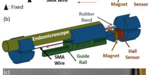
Fiber-optic endomicroscopes open new avenues for the application of non-linear optics to novel in vivo applications. To achieve focus scanning in vivo, shape memory alloy (SMA) wires have been used to move optical elements in miniature endomicroscopes. However, this method has various limitations, making it difficult to achieve accurate and reliable depth scanning. Here we present a feedback-controlled SMA depth scanner. With a Hall effect sensor, contraction of the SMA wire can be tracked in real time, rendering accurate and robust control of motion. The SMA depth scanner can achieve up to 490 µm travel and with open-loop operation, it can move more than 350 µm within one second. With the feedback loop engaged, submicron positioning accuracy was achieved along with superior positioning stability. The high-precision positioning capability of the SMA depth scanner was verified by depth-resolved nonlinear endomicroscopic imaging of mouse brain samples.

Congratulations! Way to go, Cody! According to Hopkins News (21 NSF Graduate Fellows to Conduct Research at Johns Hopkins): Cody is interested in how the brain’s glial cells modulate neural …
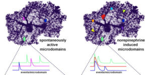
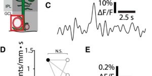
Astrocytes extend highly branched processes that form functionally isolated microdomains, facilitating local homeostasis by redistributing ions, removing neurotransmitters, and releasing factors to influence blood flow and neuronal activity. Microdomains exhibit spontaneous increases in calcium (Ca2+), but the mechanisms and functional significance of this localized signaling are unknown. By developing conditional, membrane-anchored GCaMP3 mice, we found that microdomain activity that occurs in the absence of inositol triphosphate (IP3)-dependent release from endoplasmic reticulum arises through Ca2+ efflux from mitochondria during brief openings of the mitochondrial permeability transition pore. These microdomain Ca2+ transients were facilitated by the production of reactive oxygen species during oxidative phosphorylation and were enhanced by expression of a mutant form of superoxide dismutase 1 (SOD1 G93A) that causes astrocyte dysfunction and neurodegeneration in amyotrophic lateral sclerosis (ALS). By localizing mitochondria to microdomains, astrocytes ensure local metabolic support for energetically demanding processes and enable coupling between metabolic demand and Ca2+ signaling events.
Interneurons are critical for proper neural network function and can activate Ca2+ signaling in astrocytes. However, the impact of the interneuron-astrocyte signaling into neuronal network operation remains unknown. Using the simplest hippocampal Astrocyte-Neuron network, i.e., GABAergic interneuron, pyramidal neuron, single CA3-CA1 glutamatergic synapse, and astrocytes, we found that interneuron-astrocyte signaling dynamically affected excitatory neurotransmission in an activity- and time-dependent manner, and determined the sign (inhibition vs potentiation) of the GABA-mediated effects. While synaptic inhibition was mediated by GABAA receptors, potentiation involved astrocyte GABAB receptors, astrocytic glutamate release, and presynaptic metabotropic glutamate receptors. Using conditional astrocyte-specific GABAB receptor (Gabbr1) knockout mice, we confirmed the glial source of the interneuron-induced potentiation, and demonstrated the involvement of astrocytes in hippocampal theta and gamma oscillations in vivo. Therefore, astrocytes decode interneuron activity and transform inhibitory into excitatory signals, contributing to the emergence of novel network properties resulting from the interneuron-astrocyte interplay.
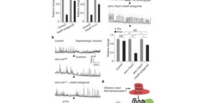
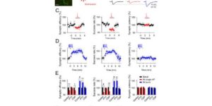
Astrocytes associate with synapses throughout the brain and express receptors for neurotransmitters that can increase intracellular calcium (Ca2+). Astrocytic Ca2+ signalling has been proposed to modulate neural circuit activity, but the pathways that regulate these events are poorly defined and in vivo evidence linking changes in astrocyte Ca2+ levels to alterations in neurotransmission or behaviour is limited. Here we show that Drosophila astrocytes exhibit activity-regulated Ca2+ signalling in vivo. Tyramine and octopamine released from neurons expressing tyrosine decarboxylase 2 (Tdc2) signal directly to astrocytes to stimulate Ca2+ increases through the octopamine/tyramine receptor (Oct-TyrR) and the transient receptor potential (TRP) channel Water witch (Wtrw), and astrocytes in turn modulate downstream dopaminergic neurons. Application of tyramine or octopamine to live preparations silenced dopaminergic neurons and this inhibition required astrocytic Oct-TyrR and Wtrw. Increasing astrocyte Ca2+ signalling was sufficient to silence dopaminergic neuron activity, which was mediated by astrocyte endocytic function and adenosine receptors. Selective disruption of Oct-TyrR or Wtrw expression in astrocytes blocked astrocytic Ca2+ signalling and profoundly altered olfactory-driven chemotaxis and touch-induced startle responses. Our work identifies Oct-TyrR and Wtrw as key components of the astrocytic Ca2+ signalling machinery, provides direct evidence that octopamine- and tyramine-based neuromodulation can be mediated by astrocytes, and demonstrates that astrocytes are essential for multiple sensory-driven behaviours in Drosophila.
The brain is critically dependent on the regulation of blood flow to nourish active neurons. One widely held hypothesis of blood flow regulation holds that active neurons stimulate Ca(2+) increases in glial cells, triggering glial release of vasodilating agents. This hypothesis has been challenged, as arteriole dilation can occur in the absence of glial Ca(2+) signaling. We address this controversy by imaging glial Ca(2+) signaling and vessel dilation in the mouse retina. We find that sensory stimulation results in Ca(2+) increases in the glial endfeet contacting capillaries, but not arterioles, and that capillary dilations often follow spontaneous Ca(2+) signaling. In IP3R2(-/-) mice, where glial Ca(2+) signaling is reduced, light-evoked capillary, but not arteriole, dilation is abolished. The results show that, independent of arterioles, capillaries actively dilate and regulate blood flow. Furthermore, the results demonstrate that glial Ca(2+) signaling regulates capillary but not arteriole blood flow.
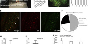
Primary sensory neurons in the DRG play an essential role in initiating pain by detecting painful stimuli in the periphery. Tissue injury can sensitize DRG neurons, causing heightened pain sensitivity, often leading to chronic pain. Despite the functional importance, how DRG neurons function at a population level is unclear due to the lack of suitable tools. Here we developed an imaging technique that allowed us to simultaneously monitor the activities of >1,600 neurons/DRG in live mice and discovered a striking neuronal coupling phenomenon that adjacent neurons tend to activate together following tissue injury. This coupled activation occurs among various neurons and is mediated by an injury-induced upregulation of gap junctions in glial cells surrounding DRG neurons. Blocking gap junctions attenuated neuronal coupling and mechanical hyperalgesia. Therefore, neuronal coupling represents a new form of neuronal plasticity in the DRG and contributes to pain hypersensitivity by “hijacking” neighboring neurons through gap junctions.
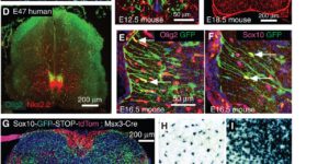
Oligodendrocyte precursor cells (OPCs) originate in the ventricular zones (VZs) of the brain and spinal cord and migrate throughout the developing central nervous system (CNS) before differentiating into myelinating oligodendrocytes (OLs). It is not known whether OPCs or OLs from different parts of the VZ are functionally distinct. OPCs persist in the postnatal CNS, where they continue to divide and generate myelinating OLs at a decreasing rate throughout adult life in rodents. Adult OPCs respond to injury or disease by accelerating their cell cycle and increasing production of OLs to replace lost myelin. They also form synapses with unmyelinated axons and respond to electrical activity in those axons by generating more OLs and myelin locally. This experience-dependent “adaptive” myelination is important in some forms of plasticity and learning, for example, motor learning. We review the control of OL lineage development, including OL population dynamics and adaptive myelination in the adult CNS.
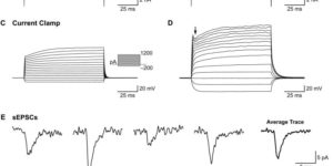
NG2(+) glial cells are a dynamic population of non-neuronal cells that give rise to myelinating oligodendrocytes in the central nervous system. These cells express numerous ion channels and neurotransmitter receptors, which endow them with a complex electrophysiological profile that is unique among glial cells. Despite extensive analysis of the electrophysiological properties of these cells, relatively little was known about the molecular identity of the channels and receptors that they express. The generation of new RNA-Seq datasets for NG2(+) cells has provided the means to explore how distinct genes contribute to the physiological properties of these progenitors. In this review, we systematically compare the results obtained through RNA-Seq transcriptional analysis of purified NG2(+) cells to previous physiological and molecular studies of these cells to define the complement of ion channels and neurotransmitter receptors expressed by NG2(+) cells in the mammalian brain and discuss the potential significance of the unique physiological properties of these cells.