Neuromodulators signal through astrocytes to alter neural circuit activity and behaviour.

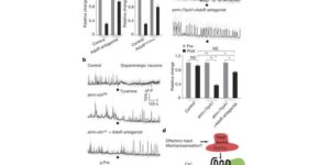
Astrocytes associate with synapses throughout the brain and express receptors for neurotransmitters that can increase intracellular calcium (Ca2+). Astrocytic Ca2+ signalling has been proposed to modulate neural circuit activity, but the pathways that regulate these events are poorly defined and in vivo evidence linking changes in astrocyte Ca2+ levels to alterations in neurotransmission or behaviour is limited. Here we show that Drosophila astrocytes exhibit activity-regulated Ca2+ signalling in vivo. Tyramine and octopamine released from neurons expressing tyrosine decarboxylase 2 (Tdc2) signal directly to astrocytes to stimulate Ca2+ increases through the octopamine/tyramine receptor (Oct-TyrR) and the transient receptor potential (TRP) channel Water witch (Wtrw), and astrocytes in turn modulate downstream dopaminergic neurons. Application of tyramine or octopamine to live preparations silenced dopaminergic neurons and this inhibition required astrocytic Oct-TyrR and Wtrw. Increasing astrocyte Ca2+ signalling was sufficient to silence dopaminergic neuron activity, which was mediated by astrocyte endocytic function and adenosine receptors. Selective disruption of Oct-TyrR or Wtrw expression in astrocytes blocked astrocytic Ca2+ signalling and profoundly altered olfactory-driven chemotaxis and touch-induced startle responses. Our work identifies Oct-TyrR and Wtrw as key components of the astrocytic Ca2+ signalling machinery, provides direct evidence that octopamine- and tyramine-based neuromodulation can be mediated by astrocytes, and demonstrates that astrocytes are essential for multiple sensory-driven behaviours in Drosophila.

Glial Cell Calcium Signaling Mediates Capillary Regulation of Blood Flow in the Retina.

The brain is critically dependent on the regulation of blood flow to nourish active neurons. One widely held hypothesis of blood flow regulation holds that active neurons stimulate Ca(2+) increases in glial cells, triggering glial release of vasodilating agents. This hypothesis has been challenged, as arteriole dilation can occur in the absence of glial Ca(2+) signaling. We address this controversy by imaging glial Ca(2+) signaling and vessel dilation in the mouse retina. We find that sensory stimulation results in Ca(2+) increases in the glial endfeet contacting capillaries, but not arterioles, and that capillary dilations often follow spontaneous Ca(2+) signaling. In IP3R2(-/-) mice, where glial Ca(2+) signaling is reduced, light-evoked capillary, but not arteriole, dilation is abolished. The results show that, independent of arterioles, capillaries actively dilate and regulate blood flow. Furthermore, the results demonstrate that glial Ca(2+) signaling regulates capillary but not arteriole blood flow.

Coupled Activation of Primary Sensory Neurons Contributes to Chronic Pain.

Primary sensory neurons in the DRG play an essential role in initiating pain by detecting painful stimuli in the periphery. Tissue injury can sensitize DRG neurons, causing heightened pain sensitivity, often leading to chronic pain. Despite the functional importance, how DRG neurons function at a population level is unclear due to the lack of suitable tools. Here we developed an imaging technique that allowed us to simultaneously monitor the activities of >1,600 neurons/DRG in live mice and discovered a striking neuronal coupling phenomenon that adjacent neurons tend to activate together following tissue injury. This coupled activation occurs among various neurons and is mediated by an injury-induced upregulation of gap junctions in glial cells surrounding DRG neurons. Blocking gap junctions attenuated neuronal coupling and mechanical hyperalgesia. Therefore, neuronal coupling represents a new form of neuronal plasticity in the DRG and contributes to pain hypersensitivity by “hijacking” neighboring neurons through gap junctions.

Oligodendrocyte Development and Plasticity.

Oligodendrocyte precursor cells (OPCs) originate in the ventricular zones (VZs) of the brain and spinal cord and migrate throughout the developing central nervous system (CNS) before differentiating into myelinating oligodendrocytes (OLs). It is not known whether OPCs or OLs from different parts of the VZ are functionally distinct. OPCs persist in the postnatal CNS, where they continue to divide and generate myelinating OLs at a decreasing rate throughout adult life in rodents. Adult OPCs respond to injury or disease by accelerating their cell cycle and increasing production of OLs to replace lost myelin. They also form synapses with unmyelinated axons and respond to electrical activity in those axons by generating more OLs and myelin locally. This experience-dependent “adaptive” myelination is important in some forms of plasticity and learning, for example, motor learning. We review the control of OL lineage development, including OL population dynamics and adaptive myelination in the adult CNS.

Electrophysiological properties of NG2(+) cells: Matching physiological studies with gene expression profiles.

NG2(+) glial cells are a dynamic population of non-neuronal cells that give rise to myelinating oligodendrocytes in the central nervous system. These cells express numerous ion channels and neurotransmitter receptors, which endow them with a complex electrophysiological profile that is unique among glial cells. Despite extensive analysis of the electrophysiological properties of these cells, relatively little was known about the molecular identity of the channels and receptors that they express. The generation of new RNA-Seq datasets for NG2(+) cells has provided the means to explore how distinct genes contribute to the physiological properties of these progenitors. In this review, we systematically compare the results obtained through RNA-Seq transcriptional analysis of purified NG2(+) cells to previous physiological and molecular studies of these cells to define the complement of ion channels and neurotransmitter receptors expressed by NG2(+) cells in the mammalian brain and discuss the potential significance of the unique physiological properties of these cells.

NMDA Receptors Enhance Spontaneous Activity and Promote Neuronal Survival in the Developing Cochlea.

Spontaneous bursts of activity in developing sensory pathways promote maturation of neurons, refinement of neuronal connections, and assembly of appropriate functional networks. In the developing auditory system, inner hair cells (IHCs) spontaneously fire Ca(2+) spikes, each of which is transformed into a mini-burst of action potentials in spiral ganglion neurons (SGNs). Here we show that NMDARs are expressed in SGN dendritic terminals and play a critical role during transmission of activity from IHCs to SGNs before hearing onset. NMDAR activation enhances glutamate-mediated Ca(2+) influx at dendritic terminals, promotes repetitive firing of individual SGNs in response to each synaptic event, and enhances coincident activity of neighboring SGNs that will eventually encode similar frequencies of sound. Loss of NMDAR signaling from SGNs reduced their survival both in vivo and in vitro, revealing that spontaneous activity in the prehearing cochlea promotes maturation of auditory circuitry through periodic activation of NMDARs in SGNs.

Spontaneous Activity of Cochlear Hair Cells Triggered by Fluid Secretion Mechanism in Adjacent Support Cells.

Spontaneous electrical activity of neurons in developing sensory systems promotes their maturation and proper connectivity. In the auditory system, spontaneous activity of cochlear inner hair cells (IHCs) is initiated by the release of ATP from glia-like inner supporting cells (ISCs), facilitating maturation of central pathways before hearing onset. Here, we find that ATP stimulates purinergic autoreceptors in ISCs, triggering Cl(-) efflux and osmotic cell shrinkage by opening TMEM16A Ca(2+)-activated Cl(-) channels. Release of Cl(-) from ISCs also forces K(+) efflux, causing transient depolarization of IHCs near ATP release sites. Genetic deletion of TMEM16A markedly reduces the spontaneous activity of IHCs and spiral ganglion neurons in the developing cochlea and prevents ATP-dependent shrinkage of supporting cells. These results indicate that supporting cells in the developing cochlea have adapted a pathway used for fluid secretion in other organs to induce periodic excitation of hair cells.

Neuron-glia Signaling in Developing Retina Mediated by Neurotransmitter Spillover.

Neuron-glia interactions play a critical role in the maturation of neural circuits; however, little is known about the pathways that mediate their communication in the developing CNS. We investigated neuron-glia signaling in the developing retina, where we demonstrate that retinal waves reliably induce calcium transients in Müller glial cells (MCs). During cholinergic waves, MC calcium transients were blocked by muscarinic acetylcholine receptor antagonists, whereas during glutamatergic waves, MC calcium transients were inhibited by ionotropic glutamate receptor antagonists, indicating that the responsiveness of MCs changes to match the neurotransmitter used to support retinal waves. Using an optical glutamate sensor we show that the decline in MC calcium transients is caused by a reduction in the amount of glutamate reaching MCs. Together, these studies indicate that neurons and MCs exhibit correlated activity during a critical period of retinal maturation that is enabled by neurotransmitter spillover from retinal synapses.

Spontaneous activity in the developing auditory system.

Spontaneous electrical activity is a common feature of sensory systems during early development. This sensory-independent neuronal activity has been implicated in promoting their survival and maturation, as well as growth and refinement of their projections to yield circuits that can rapidly extract information about the external world. Periodic bursts of action potentials occur in auditory neurons of mammals before hearing onset. This activity is induced by inner hair cells (IHCs) within the developing cochlea, which establish functional connections with spiral ganglion neurons (SGNs) several weeks before they are capable of detecting external sounds. During this pre-hearing period, IHCs fire periodic bursts of Ca(2+) action potentials that excite SGNs, triggering brief but intense periods of activity that pass through auditory centers of the brain. Although spontaneous activity requires input from IHCs, there is ongoing debate about whether IHCs are intrinsically active and their firing periodically interrupted by external inhibitory input (IHC-inhibition model), or are intrinsically silent and their firing periodically promoted by an external excitatory stimulus (IHC-excitation model). There is accumulating evidence that inner supporting cells in Kölliker’s organ spontaneously release ATP during this time, which can induce bursts of Ca(2+) spikes in IHCs that recapitulate many features of auditory neuron activity observed in vivo. Nevertheless, the role of supporting cells in this process remains to be established in vivo. A greater understanding of the molecular mechanisms responsible for generating IHC activity in the developing cochlea will help reveal how these events contribute to the maturation of nascent auditory circuits.

Large-scale recording of astrocyte activity.

Astrocytes are highly ramified glial cells found throughout the central nervous system (CNS). They express a variety of neurotransmitter receptors that can induce widespread chemical excitation, placing these cells in an optimal position to exert global effects on brain physiology. However, the activity patterns of only a small fraction of astrocytes have been examined and techniques to manipulate their behavior are limited. As a result, little is known about how astrocytes modulate CNS function on synaptic, microcircuit, or systems levels. Here, we review current and emerging approaches for visualizing and manipulating astrocyte activity in vivo. Deciphering how astrocyte network activity is controlled in different physiological and pathological contexts is crucial for defining their roles in the healthy and diseased CNS.