Synaptic signaling between neurons and glia.

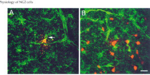
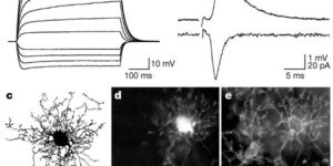
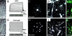
Rapid signaling between vertebrate neurons occurs primarily at synapses, intercellular junctions where quantal release of neurotransmitter triggers rapid changes in membrane conductance through activation of ionotropic receptors. Glial cells express many of these same ionotropic receptors, yet little is known about how receptors in glial cells become activated in situ. Because synapses were thought to be the sole provenance of neurons, it has been assumed that these receptors must be activated following diffusion of transmitter out of the synaptic cleft, or through nonsynaptic mechanisms such as transporter reversal. Two recent reports show that a ubiquitous class of progenitors that express the proteoglycan NG2 (NG2 cells) engage in rapid signaling with glutamatergic and gamma-aminobutyric acid (GABA)ergic neurons through direct neuron-glia synapses. Quantal release of transmitter from neurons at these sites triggers rapid activation of aminomethylisoxazole propionic acid (AMPA) or GABA(A) receptors in NG2 cells. These currents exhibit properties consistent with direct rather than spillover-mediated transmission, and electron micrographic analyses indicate that nerve terminals containing clusters of synaptic vesicles form discrete junctions with NG2 cell processes. Although activation of AMPA or GABA(A) receptors depolarize NG2 cells, these receptors are more likely to serve as routes for ion flux rather than as current sources for depolarization, because the amplitudes of the synaptic transients are small and the resting membrane potential of NG2 cells is highly negative. The ability of both glutamate and GABA to influence the morphology, physiology, and development of NG2 cells in vitro suggests that this rapid form of signaling may play important roles in adapting the behavior of these cells to the needs of surrounding neurons in vivo.

Synaptic signaling between GABAergic interneurons and oligodendrocyte precursor cells in the hippocampus

Oligodendrocyte precursor cells (OPCs) express receptors for many neurotransmitters, but the mechanisms responsible for their activation are poorly understood. We have found that quantal release of GABA from interneurons elicits GABA(A) receptor currents with rapid rise times in hippocampal OPCs. These currents did not exhibit properties of spillover transmission or release by transporters, and immunofluorescence and electron microscopy suggest that interneuronal terminals are in direct contact with OPCs, indicating that these GABA currents are generated at direct interneuron-OPC synapses. The reversal potential of OPC GABA(A) currents was -43 mV, and interneuronal firing was correlated with transient depolarizations induced by GABA(A) receptors; however, GABA application induced a transient inhibition of currents mediated by AMPA receptors in OPCs. These results indicate that OPCs are a direct target of interneuronal collaterals and that the GABA-induced Cl(-) flux generated by these events may influence oligodendrocyte development by regulating the efficacy of glutamatergic signaling in OPCs.

Physiological characteristics of NG2-expressing glial cells.

Antibodies against the chondroitin sulfate proteoglycan NG2 label a subpopulation of glial cells within the CNS, which have a small cell body and thin radiating processes. Physiological recordings from these small cells in acute brain slices have revealed that they possess unique properties, suggesting that they may comprise a class of glial cells distinct from astrocytes, oligodendrocytes, or microglia. NG2-expressing glial cells (abbreviated as “NG2 cells” here) have a moderate input resistance and are not dye- or tracer-coupled to adjacent cells. They express voltage-gated Na+, K+ and Ca2+ conductances, though they do not exhibit regenerative Na+ or Ca2+ action potentials due to the much larger K+ conductances present. In addition to voltage-gated conductances, they express receptors for various neurotransmitters. In the hippocampus, AMPA and GABAA receptors on these cells are activated by release of transmitter from neurons at defined synaptic junctions that are formed with CA3 pyramidal neurons and GABAergic interneurons. These rapid forms of neuron-glial communication may regulate the proliferation rate of NG2 cells or their development into mature oligodendrocytes. These depolarizing inputs may also trigger the release of neuroactive substances from NG2 cells, providing feedback regulation of signaling at neuronal synapses. Although the presence of Ca2+ permeable AMPA receptors provides a pathway to link neuronal activity to Ca2+ dependent processes within the NG2 cells, these receptors also put these cells at risk for glutamate-associated excitotoxicity. This vulnerability to the sustained elevation of glutamate may underlie ischemic induced damage to white matter tracts and contribute to cerebral palsy in premature infants.

Glutamatergic synapses on oligodendrocyte precursor cells in the hippocampus.

Fast excitatory neurotransmission in the central nervous system occurs at specialized synaptic junctions between neurons, where a high concentration of glutamate directly activates receptor channels. Low-affinity AMPA (alpha-amino-3-hydroxy-5-methyl isoxazole propionic acid) and kainate glutamate receptors are also expressed by some glial cells, including oligodendrocyte precursor cells (OPCs). However, the conditions that result in activation of glutamate receptors on these non-neuronal cells are not known. Here we report that stimulation of excitatory axons in the hippocampus elicits inward currents in OPCs that are mediated by AMPA receptors. The quantal nature of these responses and their rapid kinetics indicate that they are produced by the exocytosis of vesicles filled with glutamate directly opposite these receptors. Some of these AMPA receptors are permeable to calcium ions, providing a link between axonal activity and internal calcium levels in OPCs. Electron microscopic analysis revealed that vesicle-filled axon terminals make synaptic junctions with the processes of OPCs in both the young and adult hippocampus. These results demonstrate the existence of a rapid signalling pathway from pyramidal neurons to OPCs in the mammalian hippocampus that is mediated by excitatory, glutamatergic synapses.